. 2019 Dec 29;12(1):125.
doi: 10.1186/s13048-019-0601-6.

Kallistatin inhibits tumour progression and platinum resistance in high-grade serous ovarian cancer

Affiliations

Kallistatin inhibits tumour progression and platinum resistance in high-grade serous ovarian cancer

Huan Wu et al. J Ovarian Res. .

Erratum in

Abstract

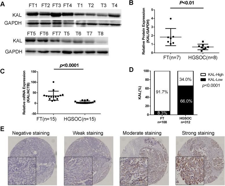
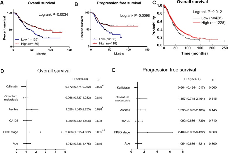
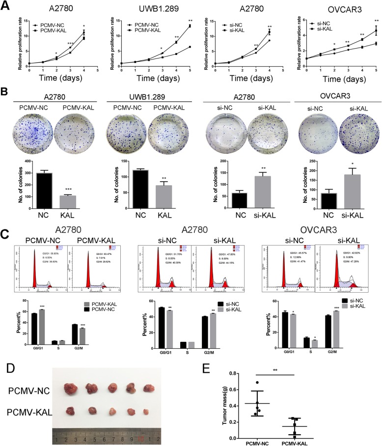
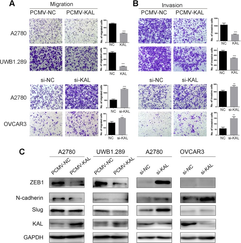
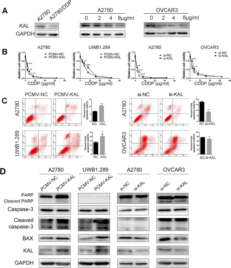
Ovarian cancer is the most lethal gynaecologic malignancy. Although there are various subtypes of ovarian cancer, high-grade serous ovarian cancer (HGSOC) accounts for 70% of ovarian cancer deaths. Chemoresistance is the primary reason for the unfavourable prognosis of HGSOC. Kallistatin (KAL), also known as SERPINA4, is part of the serpin family. Kallistatin has been discovered to exert multiple effects on angiogenesis, inflammation and tumour progression. However, the roles and clinical significance of kallistatin in HGSOC remain unclear. Here, we showed that kallistatin was significantly downregulated in HGSOC compared to normal fallopian tube (FT) tissues. Low expression of kallistatin was associated with unfavourable prognosis and platinum resistance in HGSOC. Overexpression of kallistatin significantly inhibited proliferation and metastasis, and enhanced platinum sensitivity and apoptosis in ovarian cancer cells. Collectively, these findings demonstrate that kallistatin serves as a prognostic predictor and provide a potential therapeutic target for HGSOC.

Keywords: Apoptosis; High-grade serous ovarian cancer; Kallistatin; Metastasis; Platinum resistance; Proliferation.


Conflict of interest statement

The authors declare that they have no conflict of interest.

Figures

Fig. 1
Fig. 1
Kallistatin was downregulated in HGSOC tissues. (a, b) Kallistatin protein expression in normal fallopian tube (FT) tissues and HGSOC (T) tissues measured by western blot. (c) Kallistatin mRNA expression in 15 normal FT tissues and 15 HGSOC tissues measured by RT-PCR. (d) Kallistatin expression in normal FT tissues and HGSOC tissues measured by immunohistochemistry (IHC). (e) Representative IHC staining of kallistatin in the HGSOC TMA
Fig. 2
Fig. 2
Low expression of kallistatin predicted poor prognosis of HGSOC. (a) Overall survival rates of HGSOC patients in the low versus high kallistatin expression group. (b) Progression free survival rates of HGSOC patients in the low versus high kallistatin expression group. (c) Overall survival analysis of serous ovarian cancer patients in data from the KM plotter database. (d, e) Forest plots depicting the results of the multivariate analysis of OS and PFS assessed by the Cox proportional hazard regression model. *p < 0.05, **p < 0.01, ***p < 0.001
Fig. 3
Fig. 3
Kallistatin (KAL) inhibited the proliferation of ovarian cancer cells in vitro and in vivo. (a) The effect of kallistatin on ovarian cancer cell proliferation as measured by MTT assays; A2780 and UWB1.289 cells were transfected stably with PCMV-NC and PCMV-KAL. A2780 and OVCAR3 cells were transfected transiently with kallistatin siRNA. (b) Colony formation assays were used to measure the effect of kallistatin on A2780, UWB1.289 and OVCAR3 cell growth. (c) Cell cycle analysis of A2780 and OVCAR3 cells. (d, e) UWB1.289 cells stably transfected with PCMV-NC and PCMV-KAL were injected subcutaneously into nude female mice. The tumour weights in the PCMV-KAL group were significantly decreased compared with those in the control group. *p < 0.05, **p < 0.01, ***p < 0.001
Fig. 4
Fig. 4
Kallistatin inhibited the migration and invasion of ovarian cancer cells in vitro. (a, b) Transwell assays were performed to measure the effect of kallistatin overexpression or knockdown on the migration and invasion of A2780, UWB1.289 and OVCAR3 cells. *p < 0.05, **p < 0.01, ***p < 0.001. (c) Western blot analysis of the EMT markers ZEB1, N-cadherin and Slug
Fig. 5
Fig. 5
Kallistatin enhanced the platinum sensitivity of ovarian cancer cells. (A) Western blot analysis of kallistatin protein levels in A2780, A2780/DDP, A2780 and OVCAR3 cells treated with cisplatin at 0, 2, 4, and 8 μg/ml for 48 h. (B) Cell viability was determined using MTT in A2780, UWB1.289 and OVCAR3 cells. (C) The proportion of apoptotic cells was measured by Annexin V-FITC/PI staining and flow cytometry after cisplatin (CDDP) treatment for 24 h. (D) Western blot analysis of apoptosis-related proteins. *p < 0.05, **p < 0.01, ***p < 0.001

References

    1. Siegel RL, Miller KD, Jemal A. Cancer statistics, 2019. CA Cancer J Clin. 2019;69:7–34. doi: 10.3322/caac.21551. - DOI - PubMed
    1. Cress RD, Chen YS, Morris CR, et al. Characteristics of long-term survivors of epithelial ovarian Cancer. Obstet Gynecol. 2015;126:491–497. doi: 10.1097/AOG.0000000000000981. - DOI - PMC - PubMed
    1. Bowtell DD, Bohm S, Ahmed AA, et al. Rethinking ovarian cancer II: reducing mortality from high-grade serous ovarian cancer. Nat Rev Cancer. 2015;15:668–679. doi: 10.1038/nrc4019. - DOI - PMC - PubMed
    1. Aebi S, Castiglione M. Newly and relapsed epithelial ovarian carcinoma: ESMO clinical recommendations for diagnosis, treatment and follow-up. Ann Oncol. 2009;20(Suppl 4):21–23. - PubMed
    1. Audeh MW, Carmichael J, Penson RT, et al. Oral poly(ADP-ribose) polymerase inhibitor olaparib in patients with BRCA1 or BRCA2 mutations and recurrent ovarian cancer: a proof-of-concept trial. Lancet. 2010;376:245–251. doi: 10.1016/S0140-6736(10)60893-8. - DOI - PubMed

MeSH terms