. 2019 Dec 5:2019:8687120.
doi: 10.1155/2019/8687120. eCollection 2019.

Increased Soluble Epoxide Hydrolase in Human Gestational Tissues from Pregnancies Complicated by Acute Chorioamnionitis

Affiliations

Increased Soluble Epoxide Hydrolase in Human Gestational Tissues from Pregnancies Complicated by Acute Chorioamnionitis

Tai-Ho Hung et al. Mediators Inflamm. .

Abstract

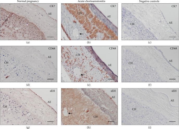
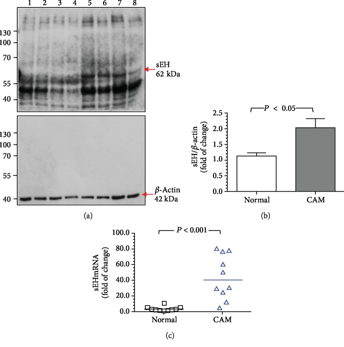
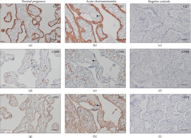
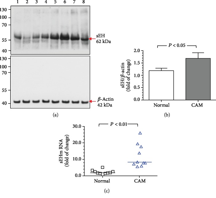
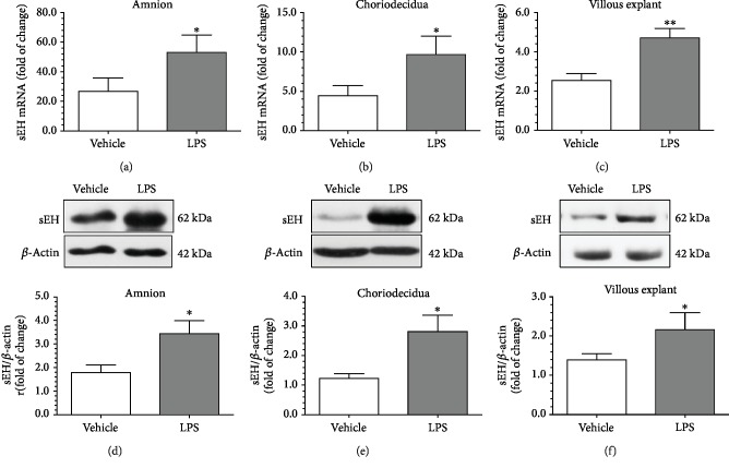
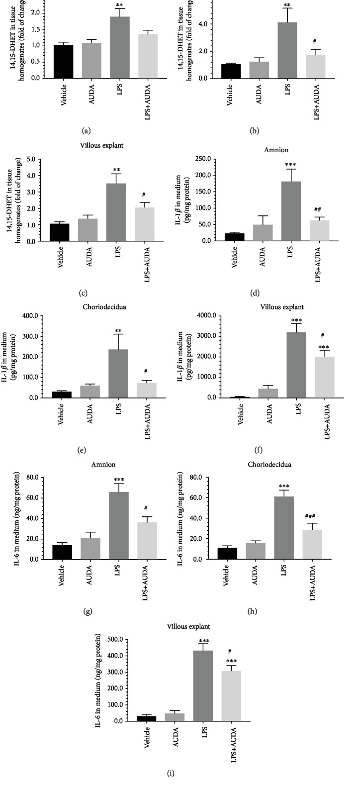
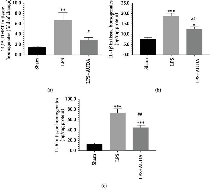
Chorioamnionitis (CAM) is primarily a polymicrobial bacterial infection involving chorionic and amniotic membranes that is associated with increased risk of preterm delivery. Epoxyeicosatrienoic acids (EETs) are eicosanoids generated from arachidonic acid by cytochrome P450 enzymes and further metabolized mainly by soluble epoxide hydrolase (sEH) to produce dihydroxyeicosatrienoic acids (DHETs). As a consequence of this metabolism of EETs, sEH reportedly exacerbates several disease states; however, its role in CAM remains unclear. The objectives of this study were to (1) determine the localization of sEH and compare the changes it undergoes in the gestational tissues (placentas and fetal membranes) of women with normal-term pregnancies and those with pregnancies complicated by acute CAM; (2) study the effects of lipopolysaccharide (LPS) on the expression of sEH in the human gestational tissues; and (3) investigate the effect of 12-(3-adamantan-1-yl-ureido)-dodecanoic acid (AUDA), a specific sEH inhibitor, on LPS-induced changes in 14,15-DHET and cytokines such as interleukin- (IL-) 1β and IL-6 in human gestational tissues in vitro and in pregnant mice. We found that women with pregnancies complicated by acute CAM had higher levels of sEH mRNA and protein in fetal membranes and villous tissues compared to those in women with normal-term pregnancies without CAM. Furthermore, fetal membrane and villous explants treated with LPS had higher tissue levels of sEH mRNA and protein and 14,15-DHET than those present in the vehicle controls, while the administration of AUDA in the media attenuated the LPS-induced production of 14,15-DHET in tissue homogenates and IL-1β and IL-6 in the media of explant cultures. Administration of AUDA also reduced the LPS-induced changes of 14,15-DHET, IL-1β, and IL-6 in the placentas of pregnant mice. Together, these results suggest that sEH participates in the inflammatory changes in human gestational tissues in pregnancies complicated by acute CAM.


Conflict of interest statement

All the authors declare that they have no conflicts of interest.

Figures

Figure 1
Figure 1
Immunoreactivity for sEH in fetal membranes from normal pregnant women and women with pregnancies complicated by acute CAM. Immunostaining of CK7 delineates the localization of amniotic epithelium and trophoblast cells in choriodecidua (a, b). Compared to normal pregnancy, fetal membranes from women with acute CAM had increased infiltration of monocytes/macrophages, as demonstrated by positive anti-CD68 immunostaining, within the amnion and choriodecidua (d, e). In normal pregnancy, immunostaining of sEH was observed in the amniotic epithelium (g). The immunostaining of sEH was generally more intense, both in the amnion and choriodecidua, in women with acute CAM compared with that in normal pregnant women (h). There was no staining observed in the negative controls when the primary antibody was substituted with nonimmune IgG (c, f, and i). Scale bar = 50 μm. AE, amniotic epithelium; CH, choriodecidua; arrows, endothelium.
Figure 2
Figure 2
The levels of sEH protein and mRNA in fetal membranes from normal pregnant women and women with pregnancies complicated by acute CAM. Compared to normal pregnant women, women with acute CAM had significantly higher levels of sEH protein (a, b) and mRNA (c) in fetal membrane homogenates. (a) Representative blots showing the expression of sEH between normal pregnant women and women with acute CAM. Lanes 1-4, fetal membrane homogenates from 4 women with normal pregnancy; lanes 5-8, homogenates from 4 women with pregnancies complicated by acute CAM. β-Actin was used to normalize loading variability. Molecular weight markers are shown at the left side of the blots. (b) Data are presented as mean ± SEM and P value is based on Student's t-test. (c) Horizontal bars represent the median values, and P value is based on the Mann-Whitney U-test. A total of 10 women with normal pregnancy and 10 women with pregnancies complicated by acute CAM were examined.
Figure 3
Figure 3
Immunoreactivity for sEH in villous tissues from normal pregnant women and women with pregnancies complicated by acute CAM. Immunostaining of CK7 delineates the localization of cytotrophoblasts and syncytiotrophoblasts (a, b). Compared to normal pregnancies, villous tissues from women with acute CAM had increased infiltration of monocytes/macrophages, as demonstrated by positive anti-CD68 immunostaining, within the stroma (d, e). In normal pregnancies, immunostaining of sEH was found mainly in the trophoblast layer and some stromal cells (g). The immunostaining of sEH was generally increased in women with acute CAM compared to normal pregnant women (h). In addition, strong sEH immunostaining was noted in the villous endothelium in women with acute CAM (arrows). There was essentially no staining at all on the negative controls when the primary antibody was substituted with nonimmune IgG (c, f, and i). Scale bar = 50 μm.
Figure 4
Figure 4
The levels of sEH protein and mRNA in villous tissues from normal pregnant women and women with pregnancies complicated by acute CAM. Compared with normal pregnant women, those with acute CAM showed significantly higher levels of sEH protein (a, b) and mRNA (c) in villous homogenates. (a) Representative blots showing the expression of sEH between normal pregnant women and those with acute CAM. Lanes 1-4, villous tissue homogenates from 4 women with normal pregnancy; lanes 5-8, homogenates from 4 women with pregnancies complicated by acute CAM. β-Actin was used to normalize loading variability. Molecular weight markers are shown at the left side of the blots. (b) Data are presented as mean ± SEM and P value is based on Student's t-test. (c) Horizontal bars represent the median values and P value is based on the Mann-Whitney U-test. A total of 10 women with normal pregnancy and 10 women with pregnancies complicated by acute CAM were examined.
Figure 5
Figure 5
LPS led to increased sEH mRNA and protein levels in fetal membrane and villous explants. Compared to the vehicle controls, the administration of LPS to the culture medium led to a significant increase in the levels of sEH mRNA and protein in the amnion and choriodecidua, respectively, with the two-chamber culture system (a, b, d, and e). Similar effects of LPS on the expression of sEH was also observed in villous explants (c and f). β-Actin was used to normalize loading variability. Data are presented as mean ± SEM, n = 10 for each group. P < 0.05 and ∗∗P < 0.01 compared with the vehicle control by Student's t-test.
Figure 6
Figure 6
Administration of AUDA reduced the levels of 14,15-DHET in tissue homogenates and the levels of IL-1β and IL-6 in the culture media of fetal membrane and villous explants treated with LPS. LPS caused a significant increase in the tissue levels of 14,15-DHET in the amnion, choriodecidua, and villous explants compared to the vehicle controls (a–c). In contrast, the administration of AUDA reduced the changes in 14,15-DHET induced by LPS; however, only the differences in choriodecidua and villous explants reached statistical significance (b, c). Similarly, the levels of IL-1β and IL-6 in the media were significantly higher in fetal membrane and villous explants stimulated with LPS than in the vehicle controls (d-i). Administration of AUDA significantly attenuated the changes in IL-1β and IL-6 in the media caused by LPS treatment in fetal membrane and villous explants. Data are presented as mean ± SEM, n = 8 for each group. ∗∗P < 0.01 and ∗∗∗P < 0.001 compared with the vehicle controls; #P < 0.05, ##P < 0.01, and ###P < 0.001 compared with explants treated with LPS, with one-way ANOVA followed by Bonferroni's post hoc test.
Figure 7
Figure 7
Administration of AUDA reduced the levels of 14,15-DHET, IL-1β, and IL-6 in the placentas of pregnant mice treated with LPS. Compared to the sham controls, pregnant mice treated with LPS had significantly higher levels of 14,15-DHET, IL-1β, and IL-6 in the placentas. Administration of AUDA significantly reduced the extent of these changes caused by LPS. Data are presented as mean ± SEM based on 8 mice for each group. ∗∗P < 0.01 and ∗∗∗P < 0.001 compared with the sham controls; #P < 0.05 and ##P < 0.01 compared with mice treated with LPS, with one-way ANOVA followed by Bonferroni's post hoc test.

Similar articles

Cited by

References

    1. Dammann O., Leviton A. Maternal intrauterine infection, cytokines, and brain damage in the preterm newborn. Pediatric Research. 1997;42(1):1–8. doi: 10.1203/00006450-199707000-00001. - DOI - PubMed
    1. Goldenberg R. L., Hauth J. C., Andrews W. W. Intrauterine infection and preterm delivery. The New England Journal of Medicine. 2000;342(20):1500–1507. doi: 10.1056/NEJM200005183422007. - DOI - PubMed
    1. Mueller-Heubach E., Rubinstein D. N., Schwarz S. S. Histologic chorioamnionitis and preterm delivery in different patient populations. Obstetrics and Gynecology. 1990;75(4):622–626. - PubMed
    1. Gaudet L. M., Smith G. N. Cerebral palsy and chorioamnionitis: the inflammatory cytokine link. Obstetrical & Gynecological Survey. 2001;56(7):433–436. doi: 10.1097/00006254-200107000-00023. - DOI - PubMed
    1. Ramsey P. S., Lieman J. M., Brumfield C. G., Carlo W. Chorioamnionitis increases neonatal morbidity in pregnancies complicated by preterm premature rupture of membranes. American Journal of Obstetrics and Gynecology. 2005;192(4):1162–1166. doi: 10.1016/j.ajog.2004.11.035. - DOI - PubMed