

Proton versus Photon Radiotherapy for Pediatric Central Nervous System Malignancies: A Systematic Review and Meta-Analysis of Dosimetric Comparison Studies
- PMID: 31885580
- PMCID: PMC6900940
- DOI: 10.1155/2019/5879723
Proton versus Photon Radiotherapy for Pediatric Central Nervous System Malignancies: A Systematic Review and Meta-Analysis of Dosimetric Comparison Studies
Abstract
Background: Radiotherapy (RT) plays a fundamental role in the treatment of pediatric central nervous system (CNS) malignancies, but its late sequelae are still a challenging question. Despite developments in modern high-conformal photon techniques and proton beam therapy (PBT) are improving the normal tissues dose-sparing while maintaining satisfactory target coverage, clinical advantages supporting the optimal treatment strategy have to be better evaluated in long-term clinical studies and assessed in further radiobiological analyses. Our analysis aimed to systematically review current knowledge on the dosimetric advantages of PBT in the considered setting, which should be the basis for future specific studies.
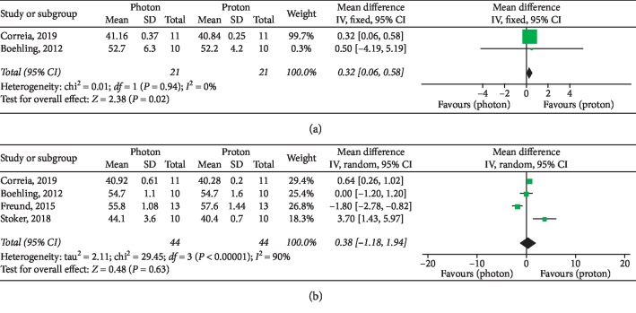
Materials and methods: A PubMed and Google Scholar search was conducted in June 2019 to select dosimetric studies comparing photon versus proton RT for pediatric patients affected by CNS tumors. Then, a systematic review and meta-analysis according to the PRISMA statement was performed. Average and standard deviation values of Conformity Index, Homogeneity Index, and mean and maximum doses to intracranial and extracranial organs at risk (OARs) were specifically evaluated for secondary dosimetric comparisons. The standardized mean differences (SMDs) for target parameters and the mean differences (MDs) for OARs were summarized in forest plots (P < 0.05 was considered statistically significant). Publication bias was also assessed by the funnel plot and Egger's regression test.
Results: Among the 88 identified papers, a total of twelve studies were included in the meta-analysis. PBT showed dosimetric advantages in target homogeneity (significant especially in the subgroup comparing PBT and 3D conformal RT), as well as in the dose sparing of almost all analyzed OARs (significantly superior results for brainstem, normal brain, and hippocampal dose constraints and for extracranial OARs parameters, excluding the kidneys). Publication bias was observed for Conformity Index.
Conclusion: Our analysis supports the evidence of dosimetric advantages of PBT over photon RT, especially in the dose sparing of normal growing tissues. Confirmations from wider well-designed studies are required.
Copyright © 2019 Roberta Carbonara et al.
Conflict of interest statement
The authors declare that they have no conflicts of interest.
Figures


References
Publication types
LinkOut - more resources
Full Text Sources
Research Materials
Miscellaneous
