

Management of Multiple Arteriovenous Malformations of the Small Bowel
- PMID: 31885600
- PMCID: PMC6927056
- DOI: 10.1155/2019/2046857
Management of Multiple Arteriovenous Malformations of the Small Bowel
Abstract
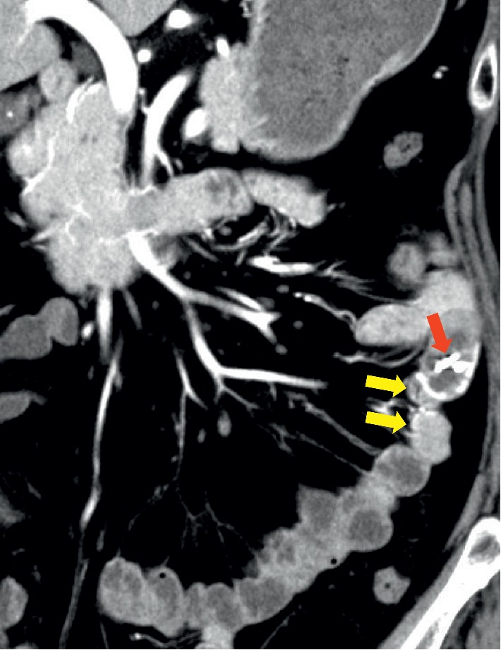
A 62-year-old Japanese female was referred to our hospital with gastrointestinal bleeding. Although small-bowel bleeding was suspected, no bleeding source was identified by enhanced computed tomography (CT), video capsule endoscopy (VCE), and double-balloon enteroscopy (DBE). Five years later, the patient had recurrent intermittent bloody stools with a significant decrease in hemoglobin levels. Although no active bleeding was observed on antegrade DBE, we detected a pulsatile submucosal uplift accompanied by a small red patch on the top of the uplift in the jejunum. Arteriovenous malformation (AVM) was suspected as the cause of small-bowel bleeding. Multiple-phase CT showed a number of small vascular ectasias during the arterial phase in the jejunum, and we confirmed the presence of multiple AVMs in the jejunum by selective angiography. To identify the location of the lesions and determine the minimal surgical margins, we performed intraoperative selective angiography with indocyanine green (ICG) injection. This technique allowed us to clearly observe the region and perform segmental small-bowel resection with minimal surgical margin. The patient reported that she has had no gastrointestinal bleeding at the two years follow-up visit.
Copyright © 2019 Masahiro Hirakawa et al.
Conflict of interest statement
The authors declare that they have no conflicts of interest.
Figures


References
-
- Longstreth G. F. Epidemiology and outcome of patients hospitalized with acute lower gastrointestinal hemorrhage: a population-based study. The American Journal of Gastroenterology. 1997;92(3):419–424. - PubMed
-
- Katz L. B. The role of surgery in occult gastrointestinal bleeding. Seminars in Gastrointestinal Disease. 1999;10(2):78–81. - PubMed
Publication types
LinkOut - more resources
Full Text Sources
