

A Method for the Isolation of Exosomes from Human and Bovine Milk
- PMID: 31885909
- PMCID: PMC6914892
- DOI: 10.1155/2019/5764740
A Method for the Isolation of Exosomes from Human and Bovine Milk
Abstract
Scope: Milk provides a natural means of nutrient supply to infants. Exosomes are an important component of milk that are not only being studied for their promise in translational medicine but also in infant nutrition. They also play important roles in intercellular communication and immune function in mammary glands and are able to transfer their materials to the recipient. Therefore, the isolation of high-quality exosomes is an important aspect of exosome research.
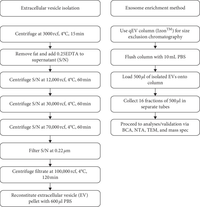
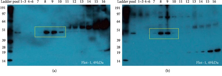
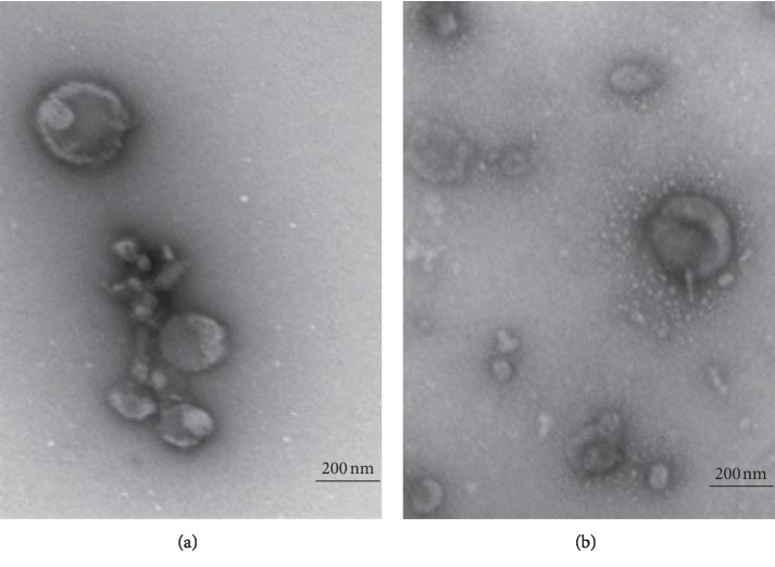
Methods and results: This study is a technical study, which provides a detailed methodology for the isolation and enrichment of exosomes from milk. In this study, we evaluate the suitability of using the exosome enrichment method that we have recently published for bovine milk, on human milk. We initially isolated extracellular vesicles from human and bovine milk on a fresh set of samples, using ultracentrifugation, and then exosomes were subsequently enriched via size exclusion chromatography (SEC). Following isolation and enrichment, exosomes from both species were characterized by particle concentration (nanoparticle tracking analysis, NTA), morphology (transmission electron microscopy, TEM), and the presence of exosomal markers (immunoblotting and mass spectrometry using information dependant acquisition (IDA)). The key exosomal characteristics of spherical/donut-shaped morphology, the presence of exosomal markers, e.g., FLOT-1 and the tetraspanins, CD9 and CD81), and particle concentration were confirmed in both human and bovine milk exosomes.
Conclusion: We conclude that our robust exosome enrichment method, previously published for bovine milk, is suitable for use on human milk.
Copyright © 2019 Kanchan Vaswani et al.
Conflict of interest statement
The authors declare no conflicts of interest.
Figures

Similar articles
-
A method for the isolation and enrichment of purified bovine milk exosomes.Reprod Biol. 2017 Dec;17(4):341-348. doi: 10.1016/j.repbio.2017.09.007. Epub 2017 Oct 10. Reprod Biol. 2017. PMID: 29030127
-
Comparison of methods for pre-processing, exosome isolation, and RNA extraction in unpasteurized bovine and human milk.PLoS One. 2021 Sep 30;16(9):e0257633. doi: 10.1371/journal.pone.0257633. eCollection 2021. PLoS One. 2021. PMID: 34591894 Free PMC article.
-
Exosome enrichment by ultracentrifugation and size exclusion chromatography.Front Biosci (Landmark Ed). 2018 Jan 1;23(5):865-874. doi: 10.2741/4621. Front Biosci (Landmark Ed). 2018. PMID: 28930577
-
Biological Activities of Extracellular Vesicles and Their Cargos from Bovine and Human Milk in Humans and Implications for Infants.J Nutr. 2017 Jan;147(1):3-10. doi: 10.3945/jn.116.238949. Epub 2016 Nov 16. J Nutr. 2017. PMID: 27852870 Free PMC article. Review.
-
A guide to mass spectrometric analysis of extracellular vesicle proteins for biomarker discovery.Mass Spectrom Rev. 2023 Mar;42(2):844-872. doi: 10.1002/mas.21749. Epub 2021 Nov 8. Mass Spectrom Rev. 2023. PMID: 34747512 Review.
Cited by
-
Extracellular vesicles: a promising cell-free therapy for cartilage repair.Future Sci OA. 2021 Dec 6;8(2):FSO774. doi: 10.2144/fsoa-2021-0096. eCollection 2022 Feb. Future Sci OA. 2021. PMID: 35070356 Free PMC article. Review.
-
Engineered exosomes for studies in tumor immunology.Immunol Rev. 2022 Nov;312(1):76-102. doi: 10.1111/imr.13107. Epub 2022 Jul 8. Immunol Rev. 2022. PMID: 35808839 Free PMC article. Review.
-
Anti-Inflammatory Potential of Cow, Donkey and Goat Milk Extracellular Vesicles as Revealed by Metabolomic Profile.Nutrients. 2020 Sep 23;12(10):2908. doi: 10.3390/nu12102908. Nutrients. 2020. PMID: 32977543 Free PMC article.
-
Nanomedicine for increasing the oral bioavailability of cancer treatments.J Nanobiotechnology. 2021 Oct 30;19(1):354. doi: 10.1186/s12951-021-01100-2. J Nanobiotechnology. 2021. PMID: 34717658 Free PMC article. Review.
-
A Review of Exosomal Isolation Methods: Is Size Exclusion Chromatography the Best Option?Int J Mol Sci. 2020 Sep 4;21(18):6466. doi: 10.3390/ijms21186466. Int J Mol Sci. 2020. PMID: 32899828 Free PMC article. Review.
References
-
- Gamez-Valero A., Monguio-Tortajada M., Carreras-Planella L., Franquesa M., Beyer K., Borras F. E. Size-exclusion chromatography-based isolation minimally alters extracellular vesicles’ characteristics compared to precipitating agents. Scientific Reports. 2016;6 doi: 10.1038/srep33641. - DOI - PMC - PubMed
-
- Koh Y. Q. Brisbane, Queensland 4029, Australia: Faculty of Medicine, The University of Queensland; 2019. Exosomes and their potential roles in dairy cow fertility. Ph.D. thesis.
LinkOut - more resources
Full Text Sources
Other Literature Sources
