

Caecocentral Scotoma: A Rare Presentation of Optic Perineuritis
- PMID: 31886042
- PMCID: PMC6901378
- DOI: 10.7759/cureus.6101
Caecocentral Scotoma: A Rare Presentation of Optic Perineuritis
Abstract
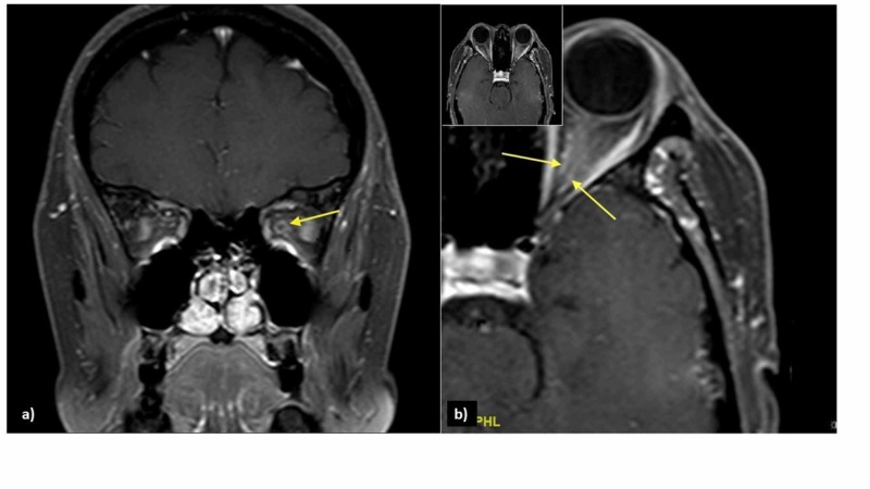
Optic perineuritis (OPN) is a subtype of optic neuritis (ON) in which the inflammatory process involves meningeal sheath surrounding the optic nerve. Clinically, OPN simulates ON. However, in contrast to ON, patient with OPN shows sparing of central vision, improves dramatically with high-dose corticosteroid, are more likely to experience recurrence after stopping treatment. We report a rare case of caecocentral scotoma observed in a female with typical ON symptoms. Her magnetic resonance imaging showed features in line with OPN. She was treated with intravenous methylprednisolone 1 g/day for five days followed by slow tapering dose of oral prednisolone for one month. Her vision improved dramatically with a resolution of visual field defect. No relapses seen within two years of follow-up.
Keywords: caecocentral scotoma; doughnut sign; optic neuritis; optic perineuritis; retrobulbar optic neuritis; tram-track sign.
Copyright © 2019, Mohamad et al.
Conflict of interest statement
The authors have declared that no competing interests exist.
Figures

References
-
- A review of optic perineuritis. Tai ELM, Tevaraj JMP, Thavaratnam LK, Mohd Noor RA, Salmah WM, Wan Hazabbah WH. Int Eye Sci. 2017;17:213–216.
-
- Optic perineuritis: clinical and radiographic features. Purvin V, Kawasaki A, Jacobson DM. Arch Ophthalmol. 2001;119:1299–1306. - PubMed
Publication types
LinkOut - more resources
Full Text Sources
Research Materials
Miscellaneous