Symptomatic Sinus Pericranii with Adult Onset Headache: A Case Report with Pathologic Perspective
- PMID: 31886152
- PMCID: PMC6911930
- DOI: 10.7461/jcen.2019.21.3.163
Symptomatic Sinus Pericranii with Adult Onset Headache: A Case Report with Pathologic Perspective
Abstract
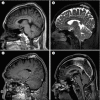
Sinus pericranii (SP) is a rare vascular anomaly of the scalp that consists of an abnormal pericranial venous channel connected to adjacent dural venous sinuses. Most SP are asymptomatic and are found in the pediatric age group. We aim to report a case of symptomatic SP in adult and describe the clinical, radiological, and pathohistological findings to help understand and differentiate this lesion from other scalp lesions. A 40-year-old man with a scalp mass was admitted to our hospital complaining of headache. The lesion enlarged when the patient was in a recumbent position or during Valsalva maneuver. The radiologic imaging suggested its diagnosis as an accessory type of SP with bone erosion. Surgical resection and cranioplasty were successfully performed, and the related headache also gradually subsided. At the 3-year follow-up, there was no recurrence on magnetic resonance imaging.
Keywords: Headache; Sinus pericranii; Vascular malformation.
© 2019 Journal of Cerebrovascular and Endovascular Neurosurgery.
Conflict of interest statement
Disclosure: The authors have no conflict of interest concerning this case report.
Figures


References
-
- Akram H, Prezerakos G, Haliasos N, O'Donovan D, Low H. Sinus pericranii: an overview and literature review of a rare cranial venous anomaly (a review of the existing literature with case examples) Neurosurg Rev. 2012 Jan;35(1):15–26. discussion 26. - PubMed
-
- Arita K, Uozumi T, Kuwabara S, Kiya K, Sumida M, Iida K. A case of scalp cavernous hemangioma simulating sinus pericranii. Hiroshima J Med Sci. 1992 Mar;41(1):19–23. - PubMed
-
- Bick DS, Brockland JJ, Scott AR. A scalp lesion with intracranial extension. Atretic cephalocele. JAMA Otolaryngol Head Neck Surg. 2015 Mar;141(3):289–290. - PubMed
-
- Gandolfo C, Krings T, Alvarez H, Ozanne A, Schaaf M, Baccin CE. Sinus pericranii: diagnostic and therapeutic considerations in 15 patients. Neuroradiology. 2007 Jun;49(6):505–514. - PubMed
-
- Guler S, Tatli B. Rare vascular pathology sinus pericranii; becomes symptomatic with pseudotumor cerebri. Turk J Pediatr. 2015 Nov-Dec;57(6):618–620. - PubMed

