. 2020 Feb 24;59(9):3523-3528.
doi: 10.1002/anie.201914411. Epub 2020 Jan 29.

A Mass-Spectrometry-Based Approach to Distinguish Annular and Specific Lipid Binding to Membrane Proteins

Affiliations

A Mass-Spectrometry-Based Approach to Distinguish Annular and Specific Lipid Binding to Membrane Proteins

Jani Reddy Bolla et al. Angew Chem Int Ed Engl. .

Abstract

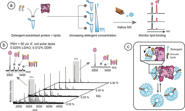
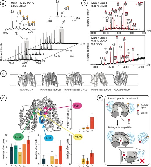
Membrane proteins engage in a variety of contacts with their surrounding lipids, but distinguishing between specifically bound lipids, and non-specific, annular interactions is a challenging problem. Applying native mass spectrometry to three membrane protein complexes with different lipid-binding properties, we explore the ability of detergents to compete with lipids bound in different environments. We show that lipids in annular positions on the presenilin homologue protease are subject to constant exchange with detergent. By contrast, detergent-resistant lipids bound at the dimer interface in the leucine transporter show decreased koff rates in molecular dynamics simulations. Turning to the lipid flippase MurJ, we find that addition of the natural substrate lipid-II results in the formation of a 1:1 protein-lipid complex, where the lipid cannot be displaced by detergent from the highly protected active site. In summary, we distinguish annular from non-annular lipids based on their exchange rates in solution.

Keywords: lipid binding; membrane protein structure; molecular dynamics; native mass spectrometry.


Conflict of interest statement

The authors declare no conflict of interest.

Figures

Figure 1
Figure 1
Detergents compete with annular lipids for interactions with PSH. a) Overview of the MS strategy employed. A solution of detergent‐solubilized protein with added lipids of interest is divided into multiple aliquots and supplemented with increasing amounts of detergent. Analysis by nMS shows a reduction of the lipid adduct peaks as a function of detergent concentration, revealing competition between lipids and detergent for binding to the protein. b) Addition of 50 μm E. coli polar lipids to PSH results in the formation of multiple lipid adducts per charge state. The 11+ charge state with lipid adducts in the presence of 0.2 % and 0.5 % NG is shown (inserts left and right, respectively). Stepwise increase of NG concentration effectively removes all bound lipids. c) Schematic representation of the competition between lipids bound at annular positions on the protein, in equilibrium with detergent molecules. The addition of excess detergent micelles dilutes the lipids, reducing binding.
Figure 2
Figure 2
CDL exhibits extended residency times at the interface of the LeuT dimer compared to annular sites. a) nMS of LeuT shows peaks indicating in mass a CDL‐mediated dimer. Incubation in 2 % NG abolishes the dimers, and lipid‐free monomers are instead detected. b) Schematic to show that LeuT forms a native lipid‐mediated dimer in the membrane. Delipidation readily removes annular lipids around the transmembrane region, whereas removal of non‐annular lipids at the LeuT interface requires a high concentration of the detergent NG (see reference 11. c) Three CDL‐binding sites on each protomer were identified in MD simulations of LeuT in a lipid bilayer. Residency times and d) k off rates of CDL molecules bound to all three sites were computed. CDL bound to residues R88/K376 at the dimer interface exhibited the slowest k off and residency times up to 1000 ns, while CDL bound to the R453/R446 and the R11/I441 site showed greater than 3‐fold faster k off rates and no lipid residency times over 200 ns. Near‐identical k off rates were observed for both halves of the dimer (Supporting Information, Figure S2).
Figure 3
Figure 3
The lipid flippase MurJ binds a single lipid‐II that does not exchange with detergent. a) Lipid adducts formed by the addition of PE to MurJ are readily exchanged by increasing the concentration of NG. The 14+ charge state of MurJ in 0.05 % LDAO with lipid adducts in the presence of 0 % and 1 % NG is shown (left and right inserts, respectively) b) The addition of lipid‐II to detergent‐solubilized MurJ results in binding of one to three lipid molecules per monomer. Increasing the concentration of OG increases the average charge of MurJ but has no pronounced effect on the binding of one lipid‐II, while all additional bound lipids are removed. c) The X‐ray crystal structures of Th. africanus MurJ in the inward, inward‐open, inward‐occluded, inward‐closed, and outward states. d) Positions of F256 (outside), R18 (active site gate), and R24 and R255 (active site) in MurJ exhibit different accessibilities for lipid substrates depending on the transport state of the protein. Monitoring contacts between UDP acyl chains and key residues in MurJ in a PE bilayer over time reveals R18, R24, and R255 in the active site are sporadically accessed by the lipid substrate in the inward state but are exposed in the outward state. e) Schematic to show how MurJ can bind multiple lipids that exchange differently with the surrounding detergent, with a single lipid‐II exhibiting much slower k off rates due to its protected location in the active site.

References

    1. Dowhan W., Bogdanov M., Biochem. Soc. Trans. 2011, 39, 767–774. - PMC - PubMed
    1. Lee A. G., Biochem. Soc. Trans. 2011, 39, 761–766. - PubMed
    1. Bechara C., Robinson C. V., J. Am. Chem. Soc. 2015, 137, 5240–5247. - PubMed
    1. Landreh M., Marty M. T., Gault J., Robinson C. V., Curr. Opin. Struct. Biol. 2016, 39, 54–60. - PMC - PubMed
    1. Palsdottir H., Hunte C., Biochim. Biophys. Acta Biomembr. 2004, 1666, 2–18. - PubMed

Publication types

LinkOut - more resources