. 2019 Dec 26;18(1):23.
doi: 10.3390/md18010023.

Quorum Quenching Properties and Probiotic Potentials of Intestinal Associated Bacteria in Asian Sea Bass Lates calcarifer

Affiliations

Quorum Quenching Properties and Probiotic Potentials of Intestinal Associated Bacteria in Asian Sea Bass Lates calcarifer

Reza Ghanei-Motlagh et al. Mar Drugs. .

Abstract

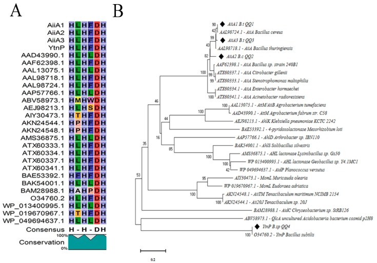
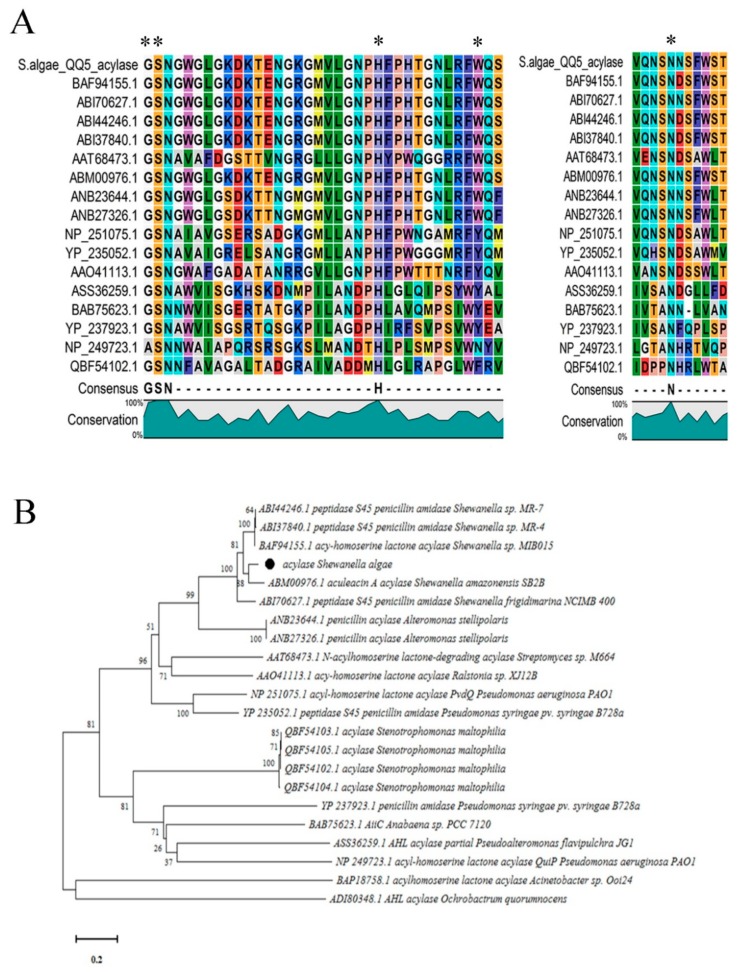
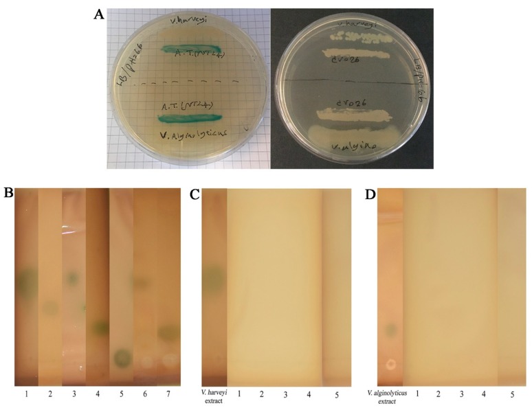
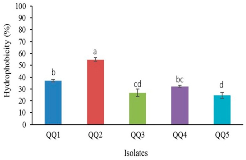
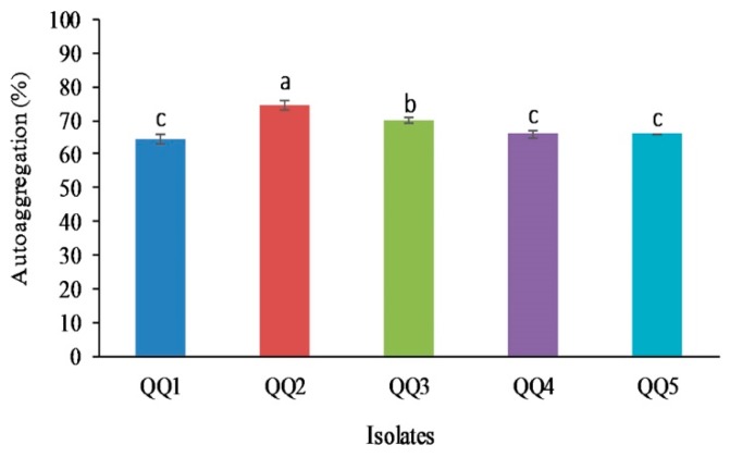
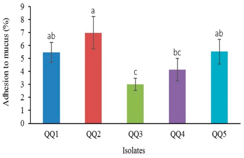
Quorum quenching (QQ), the enzymatic degradation of N-acyl homoserine lactones (AHLs), has been suggested as a promising strategy to control bacterial diseases. In this study, 10 AHL-degrading bacteria isolated from the intestine of barramundi were identified by 16S rDNA sequencing. They were able to degrade both short and long-chain AHLs associated with several pathogenic Vibrio species (spp.) in fish, including N-[(RS)-3-Hydroxybutyryl]-l-homoserine lactone (3-oh-C4-HSL), N-Hexanoyl-l-homoserine lactone (C6-HSL), N-(β-Ketocaproyl)-l-homoserine lactone (3-oxo-C6-HSL), N-(3-Oxodecanoyl)-l-homoserine lactone (3-oxo-C10-HSL), N-(3-Oxotetradecanoyl)-l-homoserine lactone (3-oxo-C14-HSL). Five QQ isolates (QQIs) belonging to the Bacillus and Shewanella genera, showed high capacity to degrade both synthetic AHLs as well as natural AHLs produced by Vibrio harveyi and Vibrio alginolyticus using the well-diffusion method and thin-layer chromatography (TLC). The genes responsible for QQ activity, including aiiA, ytnP, and aaC were also detected. Analysis of the amino acid sequences from the predicted lactonases revealed the presence of the conserved motif HxHxDH. The selected isolates were further characterized in terms of their probiotic potentials in vitro. Based on our scoring system, Bacillus thuringiensis QQ1 and Bacillus cereus QQ2 exhibited suitable probiotic characteristics, including the production of spore and exoenzymes, resistance to bile salts and pH, high potential to adhere on mucus, appropriate growth abilities, safety to barramundi, and sensitivity to antibiotics. These isolates, therefore, constitute new QQ probiotics that could be used to control vibriosis in Lates calcalifer.

Keywords: Lates calcalifer; probiotic; quorum quenching; vibriosis.


Conflict of interest statement

The authors declare that they have no competing interests.

Figures

Figure 1
Figure 1
Calibration curves between the doses of N-acyl homoserine lactones (AHLs) and the halo diameter generated by the biosensors (mean ± SD, n = 3).
Figure 2
Figure 2
Degradation of synthetic AHLs. (AE) Kinetics of AHL degradation by the selected strains. Measurements were taken at 24 (white bars) and 48 h (grey bars) intervals (mean ± SD, n = 3). (F) QQ activity detected by C. violaceum CV026 and A. tumefaciens NTL4: no AHL degradation (−), imperfect AHL degradation (+), and perfect AHL degradation (++).
Figure 3
Figure 3
Agarose gel electrophoresis showing the PCR-amplified products from the Quorum quenching (QQ) genes. (A) Amplification of the AiiA gene from B. thuringiensis QQ1 (lane 1), B. cereus QQ2 (lane 2), and B. thuringiensis QQ3 (lane 3) with a product size ~750 bp. (B) Partial amplification of the YtnP gene from Bacillus sp. QQ4 (lane 1, expected amplicon size ~530 bp). (C) Partial amplification of the acylase gene homolog from S. algae (PCR product size ~820 bp, lane 1). M stands for marker and -ve for negative control.
Figure 4
Figure 4
Comparison of the amino acid sequences of AHL lactonase enzymes. (A) Sequences corresponding to lactonase AiiA 1–3 (from QQ1, QQ2, QQ3) and YtnP (from QQ4) were compared with other known AHL lactonases from the MBL fold metallohydrolase superfamily, and all represented the common conserved motif ‘HxHxDH’. Each part of this motif is represented by a specific letter and color. Columns that are of the same color and with the same letter show motifs conserved between all sequences. (B) Phylogenetic analysis based upon amino acid sequences of detected AHL lactonases (AiiA and YtnP, black quadrates) and the homolog sequences, including AiiA from Bacillus thuringiensis (AAL98718.1), AiiA from Bacillus cereus (AAL98724.1), AiiA from Bacillus sp. strain 240B1 (AAF62398.1), AttM/AiiB from Agrobacterium tumefaciens (AAL13075.1), AhlD from Arthrobacter sp. IBN110 (AAP57766.1), QlcA from uncultured Acidobacteria bacterium cosmid p2H8 (ABV58973.1), AhlK from Klebsiella pneumoniae KCTC 2242 (AEJ98213.1), MomL from Muricauda olearia (AIY30473.1), MomL from Eudoraea adriatica (WP_019670967.1), AidC from Chryseobacterium sp. StRB126 (BAM28988.1), 4-pyridoxolactonase from Mesorhizobium loti (BAE53392.1), AhlS from Solibacillus silvestris (BAK54001.1), AHL lactonase from Lysinibacillus sp. Gs50 (AMS36875.1), AHL lactonase from Geobacillus sp. Y4.1MC1 (WP_013400995.1), AiiA from Enterobacter hormaechei (ATX60334.1), AiiA from Acinetobacter radioresistens (ATX60341.1), AiiA from Citrobacter gillenii (ATX60337.1), AiiA from Stenotrophomonas maltophilia (ATX60333.1), AttM from Agrobacterium fabrum str. C58 (AAD43990.1), AiiTM from Tenacibaculum maritimum NCIMB 2154 (AKN24548.1), Aii20J from Tenacibaculum sp. 20J (AKN24544.1), and AidP from Planococcus versutus (WP_049694637.1). The dendrogram was generated by the neighbor-joining method using the MEGA X software. The scale bar represents 0.2 substitutions per amino acid position.
Figure 5
Figure 5
Comparison of the amino acid sequences of quorum quenching acylase enzymes. (A) The comparison of amino acid sequences of putative N-acyl homoserine lactones (AHLs) acylase (from QQ5) with other known AHL acylases from the Ntn hydrolase superfamily represented the important residues for the catalytic processing in known acylases (shown by asterisks). Each part of this motif is represented by a specific letter and color. Columns that are of the same color and with the same letter show motifs conserved between all sequences. (B) Phylogenetic tree based on amino acid sequences resulting from the acylase of the Shewanella algae (Black circle) and other known acylases from Stenotrophomonas maltophilia (QBF54102.1, QBF54103.1, QBF54104.1, and QBF54105.1), Acinetobacter sp. Ooi24 (BAP18758.1), Ochrobactrum quorumnocens (ADI80348.1), Streptomyces sp. M664 (AAT68473.1), Pseudomonas aeruginosa PAO1 (NP_251075.1 and NP_249723.1), Shewanella sp. MIB015 (BAF94155.1), Alteromonas stellipolaris (ANB23644.1 and ANB27326.1), Pseudoalteromonas flavipulchra JG1 (ASS36259.1), Ralstonia sp. XJ12B (AAO41113.1), Anabaena sp. PCC 7120 (BAB75623.1), Shewanella sp. MR-7 (ABI44246.1), Shewanella sp. MR-4 (ABI37840.1), Shewanella frigidimarina NCIMB 400 (ABI70627.1), Shewanella amazonensis SB2B (ABM00976.1) and Pseudomonas syringae pv. syringae B728a (YP_235052.1 and YP_237923.1). The tree was constructed by the neighbor-joining method using the MEGA X software. The scale bar represents 0.2 substitutions per amino acid position.
Figure 6
Figure 6
Detection and degradation of natural AHLs. (A) Detection of AHLs produced by tested V. harveyi and V. alginolyticus by the cross-feeding assay. Induction of A. tumefaciens NTL4 (left plate) vs. non-induction of C. violaceum CV026 (right plate). (B) Thin-layer chromatography (TLC) analysis for tentative identification of AHLs extracted from tested V. harveyi (lane 6) and V. alginolyticus (lane 7). Synthetic AHLs were used as standards. Lane 1 (3-oh-C4-HSL), lane 2 (C6-HSL), lane 3 (3-oxo-C6-HSL), lane 4 (3-oxo-C10-HSL), and lane 5 (3-oxo-C14-HSL). (CD) Visualization of the AHL-degrading activity of selected QQIs against natural AHLs of V. harveyi and V. alginolyticus using A. tumefaciens NTL4 overlay on TLC plates. The absence of blue color development in A. tumefaciens NTL4 indicates positive QQ activity. AHLs extracted from V. harveyi and V. alginolyticus cultures were regarded as negative controls. QQ1 (lane 1), QQ2 (lane 2), QQ3 (lane 3), QQ4 (lane 4), QQ5 (lane 5).
Figure 7
Figure 7
Hydrophobicity percentage of the quorum quenching bacteria (Mean ± SD, n = 3). Dissimilar letters show a significant difference among the isolates (P < 0.05). Means were rated as a (4), ab (3.5), b (3), bc (2.5), c (2), cd (1.5), d (1). The total score for the isolates was recorded as follows: QQ1 (3), QQ2 (4), QQ3 (1.5), QQ4 (2.5), QQ5 (1).
Figure 8
Figure 8
The percentage of auto-aggregation in quorum quenching bacteria (Mean ± SD, n = 3). Different alphabets represent a significant difference between isolates (P < 0.05). The rating of means was performed as follows a (4), b (3), c (2). The total score of each isolate was QQ1 (2), QQ2 (4), QQ3 (3), QQ4 (2), QQ5 (2).
Figure 9
Figure 9
The adhesion potential of quorum quenching bacteria to intestinal mucus (Mean ± SD, n = 6). Different superscript letters denote significant differences between means (P < 0.05). Values were scored as a (4), ab (3.5), b (3), bc (2.5), c (2). The total score calculated for each isolate was the following: QQ1 (3.5), QQ2 (4), QQ3 (2), QQ4 (2.5), QQ5 (3.5).

Similar articles

Cited by

References

    1. Ina-Salwany M., Al-saari N., Mohamad A., Mursidi F.A., Mohd-Aris A., Amal M., Kasai H., Mino S., Sawabe T., Zamri-Saad M. Vibriosis in fish: A review on disease development and prevention. J. Aquat. Anim. Health. 2019;31:3–22. doi: 10.1002/aah.10045. - DOI - PubMed
    1. Dawood M.A., Koshio S., Ishikawa M., Yokoyama S., El Basuini M.F., Hossain M.S., Nhu T.H., Dossou S., Moss A.S. Effects of dietary supplementation of Lactobacillus rhamnosus or/and Lactococcus lactis on the growth, gut microbiota and immune responses of red sea bream, Pagrus major. Fish Shellfish. Immunol. 2016;49:275–285. doi: 10.1016/j.fsi.2015.12.047. - DOI - PubMed
    1. Novriadi R. Vibriosis in aquaculture. Omni-Akuatika. 2016:12. doi: 10.20884/1.oa.2016.12.1.24. - DOI
    1. Vezzulli L., Colwell R.R., Pruzzo C. Ocean warming and spread of pathogenic vibrios in the aquatic environment. Microb. Ecol. 2013;65:817–825. doi: 10.1007/s00248-012-0163-2. - DOI - PubMed
    1. Baker-Austin C., Oliver J.D., Alam M., Ali A., Waldor M.K., Qadri F., Martinez-Urtaza J. Vibrio spp. infections. Nat. Rev. Dis. Primers. 2018;4:1–19. doi: 10.1038/s41572-018-0005-8. - DOI - PubMed

LinkOut - more resources