Review
. 2019 Dec 26;9(1):69.
doi: 10.3390/cells9010069.

The Role of Cyclic AMP Signaling in Cardiac Fibrosis

Affiliations
Review

The Role of Cyclic AMP Signaling in Cardiac Fibrosis

Marion Delaunay et al. Cells. .

Abstract

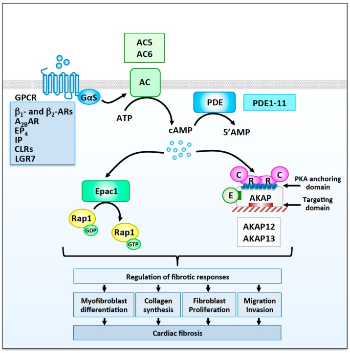
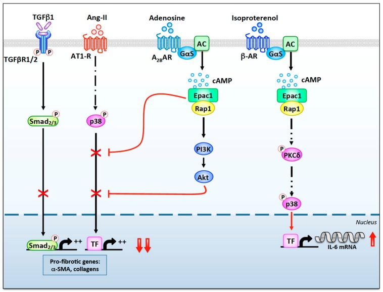
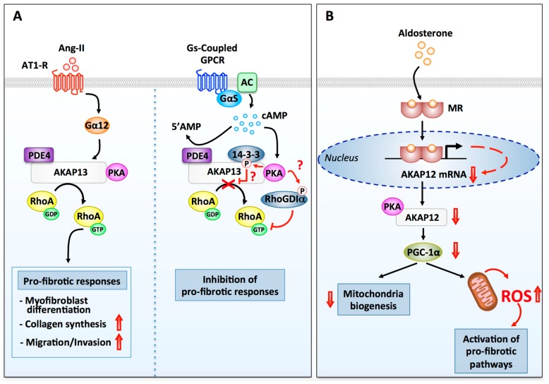
Myocardial stress and injury invariably promote remodeling of the cardiac tissue, which is associated with cardiomyocyte death and development of fibrosis. The fibrotic process is initially triggered by the differentiation of resident cardiac fibroblasts into myofibroblasts. These activated fibroblasts display increased proliferative capacity and secrete large amounts of extracellular matrix. Uncontrolled myofibroblast activation can thus promote heart stiffness, cardiac dysfunction, arrhythmias, and progression to heart failure. Despite the well-established role of myofibroblasts in mediating cardiac disease, our current knowledge on how signaling pathways promoting fibrosis are regulated and coordinated in this cell type is largely incomplete. In this respect, cyclic adenosine monophosphate (cAMP) signaling acts as a major modulator of fibrotic responses activated in fibroblasts of injured or stressed hearts. In particular, accumulating evidence now suggests that upstream cAMP modulators including G protein-coupled receptors, adenylyl cyclases (ACs), and phosphodiesterases (PDEs); downstream cAMP effectors such as protein kinase A (PKA) and the guanine nucleotide exchange factor Epac; and cAMP signaling organizers such as A-kinase anchoring proteins (AKAPs) modulate a variety of fundamental cellular processes involved in myocardial fibrosis including myofibroblast differentiation, proliferation, collagen secretion, and invasiveness. The current review will discuss recent advances highlighting the role of cAMP and AKAP-mediated signaling in regulating pathophysiological responses controlling cardiac fibrosis.

Keywords: A-kinase anchoring protein (AKAP), adenylyl cyclase; cardiac fibrosis; cardiac remodeling; cyclic AMP; phosphodiesterase; protein kinase A.


Conflict of interest statement

The authors declare no conflict of interest.

Figures

Figure 1
Figure 1
Cyclic adenosine monophosphate (cAMP) signaling modulators involved in the regulation fibrotic responses in cardiac fibroblasts. Cardiac fibroblasts express several Gs-coupled G protein-coupled receptors (GPCRs) including β1- and β2- adrenergic receptors (ARs), A2B adenosine receptors (A2BRs), prostaglandin E2 receptor 4 (EP4), prostacyclin receptors (IP), calcitonin receptor-like receptors (CLRs), and relaxin receptors (LGR7), which activate cAMP signaling cascades involved in the regulation of fibrotic responses. Stimulation of different membrane GPCRs is believed to induce the activation of distinct pool of ACs and the generation of separate cAMP microdomains. Phosphodiesterases (PDEs), which promote degradation of cAMP to AMP, are also involved in shaping intracellular cAMP microdomains. Two adenylyl cyclases, AC5 and AC6, as well as well as several phosphodiesterases families have been show to modulate cardiac fibrosis. cAMP activates two main effectors, Epac1 and protein kinase A (PKA). Epac1 catalyzes the GDP to GTP exchange on Rap1, whereas PKA, which are anchored at specific subcellular sites by A-kinase anchoring protein (AKAPs), promotes the phosphorylation of cellular substrates on serine and threonine residues. The anchoring sites for PKA and other signaling enzymes (E) as well as the AKAP targeting domains are indicated. Both Epac1 and PKA/AKAP complexes modulate, positively or negatively, multiple fibrotic responses including myofibroblast differentiation, collagen production, proliferation, migration, and invasion, which contribute to the development of myocardial fibrosis.
Figure 2
Figure 2
The anti- and pro-fibrotic roles of Epac1 in cardiac fibroblasts. Stimulation of A2BRs by adenosine leads to the local activation of Gαs and AC (AC5 and/or AC6). Generation of cAMP enhances an Epac1-dependent anti-fibrotic pathway involving Rap1, PI3K, and Akt, which inhibits TGFβR1/2-Smad2/3 and AT1-R-p38 signaling. Epac1 can also inhibit Ang-II- and TGFβ1-induced pro-fibrotic signaling through a Rap1-independent pathway. In contrast, cAMP-mediated activation of Epac1 following β-AR stimulation leads to Rap1-dependent phosphorylation of PKCδ. Once activated, PKCδ translocates to the nuclear region where it activates p38, which enhances transcription of the IL-6 gene through the activation of an uncharacterized transcription factor (TF).
Figure 3
Figure 3
Regulation of fibrotic signaling pathways by PKA and AKAPs. (A) AKAP13-dependent modulation of fibrotic responses in ventricular fibroblasts. (Left side) Stimulation of AT1-Rs by Ang-II enhances AKAP13 Rho-guanine nucleotide exchange factor (GEF) activity through a signaling pathway that involves Gα12. Active RhoA released from AKAP13 promotes several pro-fibrotic responses including myofibroblast differentiation, collagen production, TGFβ1 production, migration, and invasion. (Right side) Phosphorylation of AKAP13 by anchored PKA is known to promote 14-3-3 recruitment and inhibition of AKAP13 RhoGEF activity. PKA has also been shown to phosphorylate Rho guanine nucleotide dissociation inhibitor α (RhoGDIα), which favors its association with RhoA-GTP and RhoA inhibition. It remains to be established whether AKAP13-anchored PKA can mediate these two inhibitory responses in cardiac fibroblasts. Termination of cAMP signaling is mediated AKAP13-anchored PDE4. (B) AKAP12-dependent regulation of reactive oxygen species (ROS) production in cardiac fibroblasts. Binding of aldosterone to mineralocorticoid receptors induces their nuclear translocation and the activation of a transcriptional response that results in the downregulation of AKAP12 mRNA and protein. Suppression of AKAP12 expression leads to the downregulation of PGC-1α. This inhibits mitochondrial biogenesis and promotes the production of ROS and oxidative stress, a potent activator of pro-fibrotic signaling pathways.

References

    1. Towbin J.A., Bowles N.E. The failing heart. Nature. 2002;415:227–233. doi: 10.1038/415227a. - DOI - PubMed
    1. Xin M., Olson E.N., Bassel-Duby R. Mending broken hearts: Cardiac development as a basis for adult heart regeneration and repair. Nat. Rev. Mol. Cell Biol. 2013;14:529–541. doi: 10.1038/nrm3619. - DOI - PMC - PubMed
    1. Hill J.A., Olson E.N. Cardiac plasticity. N. Engl. J. Med. 2008;358:1370–1380. doi: 10.1056/NEJMra072139. - DOI - PubMed
    1. Barry S.P., Townsend P.A. What causes a broken heart—molecular insights into heart failure. Int. Rev. Cell Mol. Biol. 2010;284:113–179. doi: 10.1016/S1937-6448(10)84003-1. - DOI - PubMed
    1. Sun Y. Myocardial repair/remodelling following infarction: Roles of local factors. Cardiovasc. Res. 2009;81:482–490. doi: 10.1093/cvr/cvn333. - DOI - PMC - PubMed