The prognostic value of median nerve thickness in diagnosing carpal tunnel syndrome using magnetic resonance imaging: a pilot study
- PMID: 31888318
- PMCID: PMC6944367
- DOI: 10.3344/kjp.2020.33.1.54
The prognostic value of median nerve thickness in diagnosing carpal tunnel syndrome using magnetic resonance imaging: a pilot study
Abstract
Background: The median nerve cross-sectional area (MNCSA) is a useful morphological parameter for the evaluation of carpal tunnel syndrome (CTS). However, there have been limited studies investigating the anatomical basis of median nerve flattening. Thus, to evaluate the connection between median nerve flattening and CTS, we carried out a measurement of the median nerve thickness (MNT).
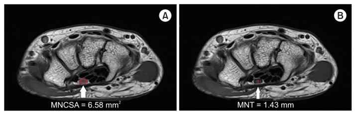
Methods: Both MNCSA and MNT measurement tools were collected from 20 patients with CTS, and from 20 control individuals who underwent carpal tunnel magnetic resonance imaging (CTMRI). We measured the MNCSA and MNT at the level of the hook of hamate on CTMRI. The MNCSA was measured on the transverse angled sections through the whole area. The MNT was measured based on the most compressed MNT.
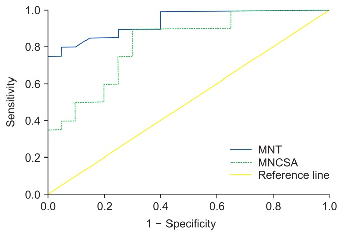
Results: The mean MNCSA was 9.01 ± 1.94 mm2 in the control group and 6.58 ± 1.75 mm2 in the CTS group. The mean MNT was 2.18 ± 0.39 mm in the control group and 1.43 ± 0.28 mm in the CTS group. Receiver operating characteristics curve analysis demonstrated that the optimal cut-off value for the MNCSA was 7.72 mm2, with 75.0% sensitivity, 75.0% specificity, and an area under the curve (AUC) of 0.82 (95% confidence interval [CI], 0.69-0.95). The best cut off-threshold of the MNT was 1.76 mm, with 85% sensitivity, 85% specificity, and an AUC of 0.94 (95% CI, 0.87-1.00).
Conclusions: Even though both MNCSA and MNT were significantly associated with CTS, MNT was identified as a more suitable measurement parameter.
Keywords: Anatomy; Area Under Curve; Carpal Tunnel Syndrome; Diagnosis; Hamate Bone; Magnetic Resonance Imaging; Median Nerve; ROC Curve; Wrist.
Conflict of interest statement
No potential conflict of interest relevant to this article was reported.
Figures
References
LinkOut - more resources
Full Text Sources
Research Materials