. 2019 Dec 30;19(1):1266.
doi: 10.1186/s12885-019-6470-y.

Gene signature characteristic of elevated stromal infiltration and activation is associated with increased risk of hematogenous and lymphatic metastasis in serous ovarian cancer

Affiliations

Gene signature characteristic of elevated stromal infiltration and activation is associated with increased risk of hematogenous and lymphatic metastasis in serous ovarian cancer

Huiran Yue et al. BMC Cancer. .

Abstract

Background: The clinical significance of hematogenous and lymphatic metastasis in ovarian cancer has been increasingly addressed, as it plays an imperative role in the formation of both intraperitoneal and distant metastases. Our objective is to identify the key molecules and biological processes potentially related to this relatively novel metastatic route in serous ovarian cancer.

Methods: Since lymphovascular space invasion (LVSI) is considered as the first step of hematogenous and lymphatic dissemination, we developed a gene signature mainly based on the transcriptome profiles with available information on LVSI status in the Cancer Genome Atlas (TCGA) dataset. We then explored the underlying biological rationale and prognostic value of the identified gene signature using multiple public databases.

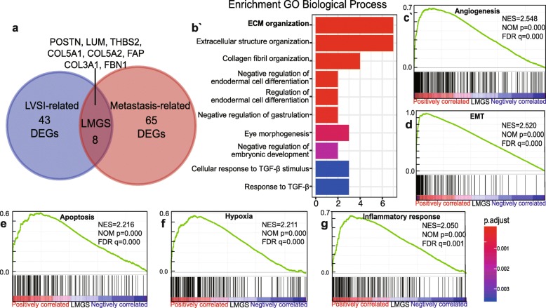
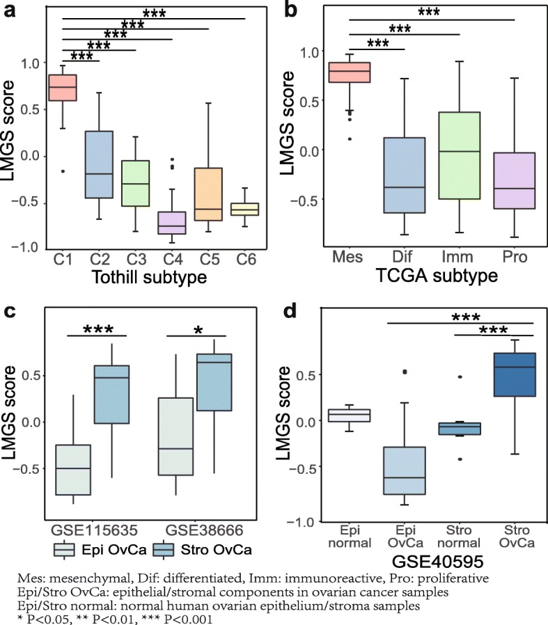
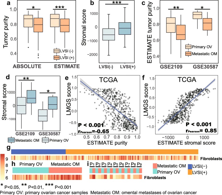
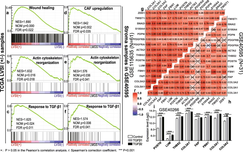
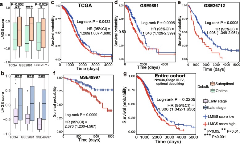
Results: We observe that primary tumors with increased risk of hematogenous and lymphatic metastasis highly express a panel of genes, namely POSTN, LUM, THBS2, COL3A1, COL5A1, COL5A2, FAP1 and FBN1. The identified geneset is characterized by enhanced deposition of extracellular matrix and extensive stromal activation. Mechanistically, both the recruitment and the activation of stromal cells, especially fibroblasts, are closely associated with lymphovascular metastasis. Survival analysis further reveals that the elevated expression of the identified genes correlates to cancer progression and poor prognosis in patients with serous ovarian cancer.

Conclusions: Our findings indicate that tumor stroma supports the hematogenous and lymphatic spread of ovarian cancer, increasing tumor invasiveness and ultimately resulting in worse survival. Thus stroma-targeted therapies may improve the clinical outcomes in combination with cytoreductive surgery and chemotherapy.

Keywords: Cancer-associated fibroblast; Hematogenous and lymphatic metastasis; Lymphovascular space invasion; Ovarian cancer; Tumor stroma.


Conflict of interest statement

The authors declare that they have no competing interests.

Figures

Fig. 1
Fig. 1
Identification and functional annotation of the gene signature associated with lymphovascular metastasis a Venn diagram showed that eight genes were common to the DEGs associated with LVSI status and the DEGs related to metastasis, representing genes potentially correlated with lymphovascular metastasis in ovarian cancer. b Functional annotation revealed that the LMGS was closely related to ECM organization. c-g Pathways correlated with cancer progression were significantly enriched in the LMGS overexpression group
Fig. 2
Fig. 2
The up-regulation of the LMGS is associated with stromal infiltration. The C1 molecular subtype of a the Tothill dataset and b TCGA mesenchymal subtype showed significant activation of the LMGS. c-d The LMGS was remarkably activated in ovarian cancer stroma in microdissected ovarian cancer samples from three independent datasets
Fig. 3
Fig. 3
LVSI-positive primary tumors resemble metastatic lesions, to which the infiltration of stromal cells contributes most. LVSI-positive samples were characterized by a reduced tumor purity and b remarkably elevated mesenchymal infiltration. Omental metastases displayed a similar trend compared to primary ovarian tumors c-d. The activation of the LMGS was e significantly negatively correlated with tumor purity and f positively correlated with mesenchymal infiltration in serous ovarian cancer samples from TCGA dataset. g The infiltration of fibroblasts was remarkably elevated in primary ovarian cancer samples with LVSI-positive status. A similar trend was observed in omental metastases compared with primary lesions in h dataset GSE2109 and was validated in i paired samples from dataset GSE30587
Fig. 4
Fig. 4
The activation of CAFs contributes to the overexpression of the LMGS in lymphovascular metastasis. a-c GSEA plots showed that pathways representative of CAF activation were significantly enriched in LVSI-positive samples, as well as in stromal profiles with high levels of the LMGS in the dataset GSE40595 d-f. The Pearson’s correlation analysis between the expression of the identified genes and CAF-specific markers was conducted based on the transcriptome profiles of ovarian cancer stroma in dataset g GSE115635 and h GSE40595. i All the identified genes were significantly unregulated in the ovarian fibroblasts NOF151-hTERT treated with either TGF-β1 or TGF-β2 compared to controls, according to the expression profiles in the dataset GSE40266
Fig. 5
Fig. 5
The LMGS up-regulation is associated with poor prognosis in patients with serous ovarian cancer. The LMGS was significantly enriched in a patients undergoing suboptimal cytoreduction and b those with late-stage disease. The prognostic significance of the LMGS in patients with serous ovarian cancer was validated in dataset c TCGA (N = 557), d GSE9891 (N = 240), e GSE26712 (N = 185), f GSE49997 (N = 171). g Survival analysis across the four validation datasets (N = 646) was conducted in patients with late-stage disease undergoing optimal cytoreduction

References

    1. Berek JS, Kehoe ST, Kumar L, Friedlander M. Cancer of the ovary, fallopian tube, and peritoneum. Int J Gynecol Obstet. 2018;143:59–78. doi: 10.1002/ijgo.12614. - DOI - PubMed
    1. Barbolina Maria. Molecular Mechanisms Regulating Organ-Specific Metastases in Epithelial Ovarian Carcinoma. Cancers. 2018;10(11):444. doi: 10.3390/cancers10110444. - DOI - PMC - PubMed
    1. Guth U, Huang DJ, Bauer G, Stieger M, Wight E, Singer G. Metastatic patterns at autopsy in patients with ovarian carcinoma. Cancer. 2007;110(6):1272–1280. doi: 10.1002/cncr.22919. - DOI - PubMed
    1. Van Berckelaer C, Brouwers AJ, Peeters DJ, Tjalma W, Trinh XB, van Dam PA. Current and future role of circulating tumor cells in patients with epithelial ovarian cancer. Eur J Surg Oncol. 2016;42(12):1772–1779. doi: 10.1016/j.ejso.2016.05.010. - DOI - PubMed
    1. Bachmann C, Bachmann R, Fend F, Wallwiener D. Incidence and impact of lymph node metastases in advanced ovarian Cancer: implications for surgical treatment. J Cancer. 2016;7(15):2241–2246. doi: 10.7150/jca.15644. - DOI - PMC - PubMed