Review
. 2019 Dec 30;14(1):12.
doi: 10.1186/s13064-019-0137-x.

Microglia in the developing retina

Affiliations
Review

Microglia in the developing retina

Fenge Li et al. Neural Dev. .

Abstract

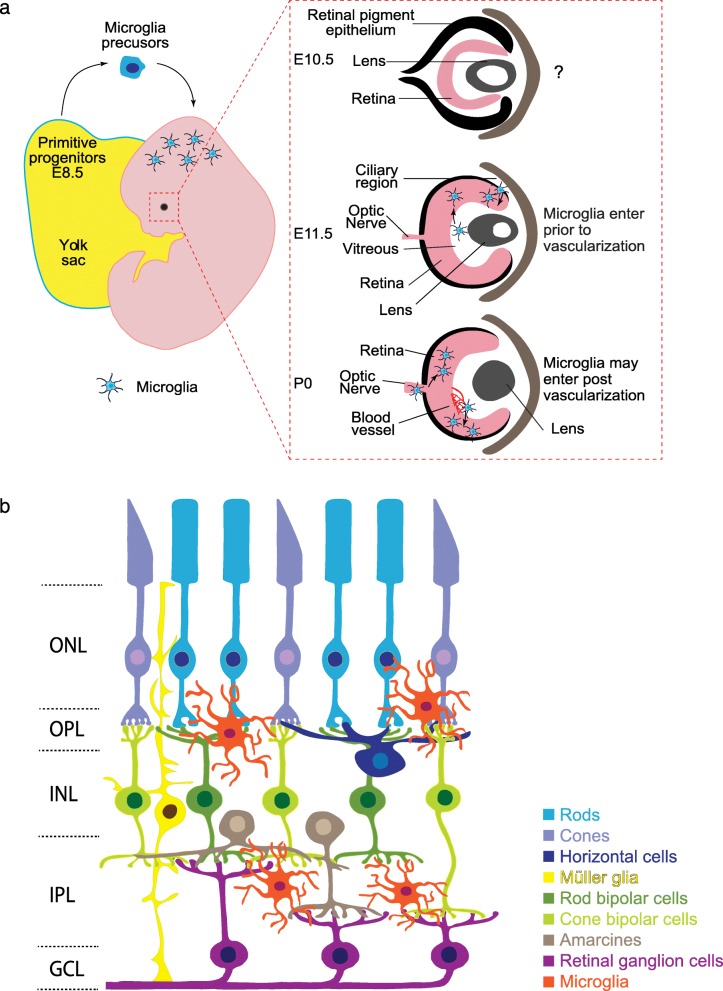
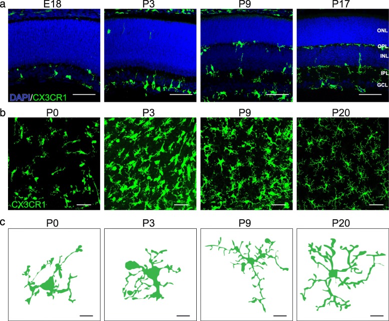
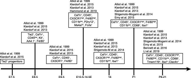
Microglia are increasingly shown to be key players in neuron development and synapse connectivity. However, the underlying mechanisms by which microglia regulate neuron function remain poorly understood in part because such analysis is challenging in the brain where neurons and synapses are intermingled and connectivity is only beginning to be mapped. Here, we discuss the features and function of microglia in the ordered mammalian retina where the laminar organization of neurons and synapses facilitates such molecular studies. We discuss microglia origins and consider the evidence for molecularly distinct microglia subpopulations and their potential for differential roles with a particular focus on the early stages of retina development. We then review the models and methods used for the study of these cells and discuss emerging data that link retina microglia to the genesis and survival of particular retina cell subtypes. We also highlight potential roles for microglia in shaping the development and organization of the vasculature and discuss cellular and molecular mechanisms involved in this process. Such insights may help resolve the mechanisms by which retinal microglia impact visual function and help guide studies of related features in brain development and disease.

Keywords: Brain; Depletion models; Development; Microglia; Retina; Synapse.


Conflict of interest statement

The authors declare no competing interests.

Figures

Fig. 1
Fig. 1
Schematic of microglia development in mouse retina. a. Timeline of microglia entry to the retina. Microglia are derived from primitive yolk sac progenitors and are thought to enter the CNS via the circulatory system. Microglia have been documented in the developing murine retina at E11.5 but may be present earlier. Two waves of retinal microglia infiltration have been proposed. The first wave occurs embryonically and may involve microglia entry through the vitreal retina surface or migration from the ciliary region. A second wave may involve microglia infiltration from the optic disc or via blood vessels. b. Schematic of the adult retina. Rod (cyan) and cone (light purple) photoreceptors reside in the outer nuclear layer (ONL) and form connections with interneurons in the outer plexiform layer (OPL). Light induced signals are then relayed to neurons in the inner nuclear layer (INL), which is comprised of horizontal cells (dark blue), Müller glia (yellow), cone and rod bipolar cells (light and dark green), and amacrines (brown). Retinal ganglion cells (magenta) receive this information through synapses in the inner plexiform layer (IPL). Their somas reside in the ganglion cell layer (GCL) along with displaced amacrine cells (not pictured). Microglia cell are found predominately in the inner retina and are largely restricted to the synaptic layers
Fig. 2
Fig. 2
Spatiotemporal distribution of microglia in the developing mouse retina. a. Representative images showing distinct spatiotemporal localization patterns of microglia across retina development (E18, P3, P9, and P17) in CX3CR1GFP/+ mice. Microglia are highly enriched at E18 and P3 in the nascent IPL where synapses are developing. At P9, microglia also become present within the developing OPL. This pattern persists into adulthood. Blue, DAPI; green, microglia. Scale bar = 50 μm. b-c. Representative images (b, scale bar = 50 μm) and single cell reconstructions (c, scale bar = 10 μm) of microglia in whole mount preparations of CX3CR1GFP/+ retina across development (P0, P3, P9, and P20). At birth, retinal microglia are amoeboid but become progressively ramified as the retina matures
Fig. 3
Fig. 3
Microglia biomarkers over CNS development. A timeline is presented that summarizes biomarkers for microglia over development in the CNS. For example, Tie2 marks microglia progenitors as early as E7.5. As microglia mature they express other markers including Csf1r, CX3CR1, and Iba1. Single-cell RNA sequencing studies have also identified potential new markers such as Tmem119, Fcrls, P2ry12, and Clec4n
Fig. 4
Fig. 4
Proposed roles of microglia in the developing retina. Microglia play important roles in phagocytosis, vascularization, and neurogenesis through distinct mechanisms during retina development. These include modulation of both hyaloid vessel regression and intraretinal vascular patterning, regulation of the numbers of astrocytes and some RGC subsets, and RPC cycling. Roles for microglia in retina synapse pruning have also been proposed, though direct evidence for these pathways awaits further study

References

    1. Schafer DP, Lehrman EK, Kautzman AG, Koyama R, Mardinly AR, Yamasaki R, et al. Microglia sculpt postnatal neural circuits in an activity and complement-dependent manner. Neuron. 2012;74(4):691–705. doi: 10.1016/j.neuron.2012.03.026. - DOI - PMC - PubMed
    1. Li Q, Barres BA. Microglia and macrophages in brain homeostasis and disease. Nat Rev Immunol. 2018;18(4):225–242. doi: 10.1038/nri.2017.125. - DOI - PubMed
    1. Colonna M, Butovsky O. Microglia function in the central nervous system during health and Neurodegeneration. Annu Rev Immunol. 2017;35:441–468. doi: 10.1146/annurev-immunol-051116-052358. - DOI - PMC - PubMed
    1. Sierra A, Abiega O, Shahraz A, Neumann H. Janus-faced microglia: beneficial and detrimental consequences of microglial phagocytosis. Front Cell Neurosci. 2013;7:6. doi: 10.3389/fncel.2013.00006. - DOI - PMC - PubMed
    1. Ashwell K. Microglia and cell death in the developing mouse cerebellum. Brain Res Dev Brain Res. 1990;55(2):219–230. doi: 10.1016/0165-3806(90)90203-B. - DOI - PubMed

Publication types

LinkOut - more resources