Case Reports
. 2019 Dec 29;12(12):e232538.
doi: 10.1136/bcr-2019-232538.

Diagnostic delay in a case of T-cell neurolymphomatosis

Affiliations
Case Reports

Diagnostic delay in a case of T-cell neurolymphomatosis

Vivien Li et al. BMJ Case Rep. .

Abstract

A 69-year-old woman presented with severe subacute painful meningoradiculoneuritis. Neurophysiology showed a patchy, proximal axonal process with widespread denervation. Cerebrospinal fluid (CSF) was lymphocytic (normal T-cell predominant) with negative cytology. MRI revealed multiple sites of enhancement, but fluorodeoxyglucose positron emission tomography was negative. Bone marrow aspirate and trephine (BMAT) showed no evidence of a lymphoproliferative condition. Right brachial plexus biopsy demonstrated mixed T-cell/B-cell endoneurial inflammation not fulfilling criteria for vasculitis. She was stabilised with high-dose steroids and cyclophosphamide, followed by mycophenolate for inflammatory myeloradiculoneuritis. However, symptoms recurred when prednisolone was weaned. Although T-cell receptor gene analysis from the initial CSF demonstrated clonal rearrangements, it was only when the same clones were identified on two repeat BMATs and CSF that T-cell neurolymphomatosis, an exceedingly rare condition, was diagnosed. This case highlights the diagnostic challenge in peripheral neurolymphomatosis related to patchy disease, variable sensitivity and specificity of investigative tools, and the influence of therapies on traditional cytological definitions of lymphoma. The clinical picture, exhaustive exclusion of alternative causes and the persistence of an abnormal T-cell clone ultimately lead to a diagnostic consensus between specialist neurology and haematology clinicians.

Keywords: haematology (incl blood transfusion); neuromuscular disease; peripheral nerve disease.


Conflict of interest statement

Competing interests: None declared.

Figures
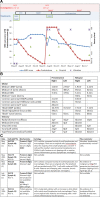
Figure 1
Figure 1
Clinical progression of disease as indicated by change in weakness (MRC sum score12) and sensory deficits over time with alterations in oral corticosteroid dose (vertical axes) and various immunomodulatory treatments (horizontal axes). Time points at which neurophysiology was performed are indicated by the red crosses and CSF by the orange crosses below the horizontal axis. (B) Neurophysiology at initial presentation (May 2015) and relapse (June 2016). (C) Serial CSF findings following initial presentation (three samples in May 2015) and at relapse (two samples in July 2016). Abnormal results are indicated in bold. BMAT, bone marrow aspirate and trephine; cm, costal margin; CMAP, compound muscle action potential; CSF, cerebrospinal fluid; CV, conduction velocity; CYC, cyclophosphamide; DML, distal motor latency; Ig, immunoglobulin; IVIg, intravenous immunoglobulin; IVMP, intravenous methylprednisolone; lymph, lymphocyte count; MMF, mycophenolate mofetil; MRC, Medical Research Council; NCS, nerve conduction study; OCB+, unpaired oligoclonal bands present in cerebrospinal fluid; RCC, red cell count; TCRg, T-cell receptor gene rearrangement; WCC, white cell count.
Figure 2
Figure 2
(A) MRI showing enhancement (as marked by red arrows) of oculomotor nerves (1), cauda equina (2) and lumbosacral plexuses on postgadolinium T1 weighted sequences (3) and enlargement and hyperintensity of the brachial plexus on T2-weighted sequences (4). (B) The cerebrospinal fluid preparation stained with Giemsa method (1) shows frequent small lymphocytes admixed with occasional intact red blood cells. Majority of the lymphocytes are CD3-positive T lymphocytes (2) with rare admixed CD20-positive B lymphocytes (3). Scale bar: 50 µm in 1–3. (C) H&E-stained section of the right brachial plexus biopsy demonstrates widespread endoneural oedema (1) and increased numbers of endoneural freely scattered and loosely arranged perivascular small lymphocytes (2). There is moderate depletion of myelinated fibres across the fascicle demonstrated by SMI94 immunostaining (3), and there is widespread active axonal degeneration with infiltration of CD68-positive macrophages (4). The endoneural inflammatory cell infiltrate is mixed, comprising CD3-positive T lymphocytes (5), CD20-positive B lymphocytes (6) and very rare, isolated CD138-positive plasma cells (not shown). Scale bar: 200 µm in A, 20 µm in B and 100 µm in 3–6. All immunostainings shown: CD3 (1:100, LN10, Leica), CD20 (1:200, L26, Leica), CD138 (1:100, MI15, DAKO) and CD68 (1:100, PG-M1, DAKO) were carried out on automated immunostainers (Roche Ventana Discovery or Leica BondMax) following the manufacturer’s guideline.

References

    1. Broccoli A, Zinzani PL. Peripheral T-cell lymphoma, not otherwise specified. Blood 2017;129:1103–12. 10.1182/blood-2016-08-692566 - DOI - PubMed
    1. Hodges E, Krishna MT, Pickard C, et al. . Diagnostic role of tests for T cell receptor (TCR) genes. J Clin Pathol 2003;56:1–11. 10.1136/jcp.56.1.1 - DOI - PMC - PubMed
    1. de Groot K, Harper L, Jayne DRW, et al. . Pulse versus daily oral cyclophosphamide for induction of remission in antineutrophil cytoplasmic antibody-associated vasculitis: a randomized trial. Ann Intern Med 2009;150:670–80. 10.7326/0003-4819-150-10-200905190-00004 - DOI - PubMed
    1. Grisariu S, Avni B, Batchelor TT, et al. . Neurolymphomatosis: an international primary CNS lymphoma Collaborative Group report. Blood 2010;115:5005–11. 10.1182/blood-2009-12-258210 - DOI - PMC - PubMed
    1. Salm LP, Van der Hiel B, Stokkel MPM. Increasing importance of 18F-FDG PET in the diagnosis of neurolymphomatosis. Nucl Med Commun 2012;33:907–16. 10.1097/MNM.0b013e3283561881 - DOI - PubMed

Publication types

MeSH terms