. 2020 Mar;61(3):316-327.
doi: 10.1194/jlr.RA119000259. Epub 2019 Dec 30.

Model systems for studying the assembly, trafficking, and secretion of apoB lipoproteins using fluorescent fusion proteins

Affiliations

Model systems for studying the assembly, trafficking, and secretion of apoB lipoproteins using fluorescent fusion proteins

Meghan T Walsh et al. J Lipid Res. 2020 Mar.

Abstract

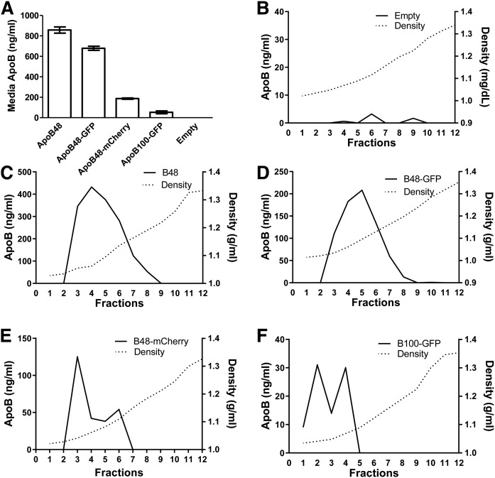
apoB exists as apoB100 and apoB48, which are mainly found in hepatic VLDLs and intestinal chylomicrons, respectively. Elevated plasma levels of apoB-containing lipoproteins (Blps) contribute to coronary artery disease, diabetes, and other cardiometabolic conditions. Studying the mechanisms that drive the assembly, intracellular trafficking, secretion, and function of Blps remains challenging. Our understanding of the intracellular and intraorganism trafficking of Blps can be greatly enhanced, however, with the availability of fusion proteins that can help visualize Blp transport within cells and between tissues. We designed three plasmids expressing human apoB fluorescent fusion proteins: apoB48-GFP, apoB100-GFP, and apoB48-mCherry. In Cos-7 cells, transiently expressed fluorescent apoB proteins colocalized with calnexin and were only secreted if cells were cotransfected with microsomal triglyceride transfer protein. The secreted apoB-fusion proteins retained the fluorescent protein and were secreted as lipoproteins with flotation densities similar to plasma HDL and LDL. In a rat hepatoma McA-RH7777 cell line, the human apoB100 fusion protein was secreted as VLDL- and LDL-sized particles, and the apoB48 fusion proteins were secreted as LDL- and HDL-sized particles. To monitor lipoprotein trafficking in vivo, the apoB48-mCherry construct was transiently expressed in zebrafish larvae and was detected throughout the liver. These experiments show that the addition of fluorescent proteins to the C terminus of apoB does not disrupt their assembly, localization, secretion, or endocytosis. The availability of fluorescently labeled apoB proteins will facilitate the exploration of the assembly, degradation, and transport of Blps and help to identify novel compounds that interfere with these processes via high-throughput screening.

Keywords: apolipoprotein B; chylomicrons; low density lipoproteins; very low density lipoproteins.


Conflict of interest statement

The authors declare that they have no conflicts of interest with the contents of this article.

Figures

Fig. 1.
Fig. 1.
Plasmids expressing different apoB fusion proteins. A: apoB48-GFP was custom-designed and constructed by Cyagen Corp. The 5′-UTR and N-terminal 48% of apoB were subcloned into the pRP.ExTri-CMV vector, which was expressed under a CMV promoter. eGFP was added after a linker sequence at the C terminus of the protein. B: apoB48-mCherry was subcloned into the pDONOR vector and then recombined into the pDEST vector using a lambda-based recombination reaction. mCherry was inserted at its C terminus. Expression was driven by the hsp70 promoter. C: apoB100-GFP synthesized by Origene. The open reading frame of full-length apoB was expressed under the CMV promoter. TurboGFP was added to the C terminus of the protein.
Fig. 2.
Fig. 2.
Fluorescent apoB fusion proteins are located in the endoplasmic reticulum and colocalize with calnexin and MTP. Cos-7 cells were plated on coverslips and transfected with plasmids expressing the various apoB constructs. A: After 48 h, cells expressing apoB48-GFP (green) were incubated with secondary antibodies conjugated to Alexa Fluor 594 (calnexin, red). B: Cells expressing apoB48-mCherry were incubated with secondary antibody conjugated to Alexa Fluor 488 (calnexin, green). C: Cos-7 cells were first transfected with plasmids expressing apoB48-GFP followed by plasmids expressing MTP-FLAG. The nucleus was stained with DAPI (blue). Cells were visualized with an Olympus confocal microscope. Images were merged to determine colocalization (yellow). The experiment was performed in duplicate, and eight adjacent fields were captured per slide. Scale bars: 10 µm.
Fig. 3.
Fig. 3.
apoB fusion proteins are secreted as lipoproteins from Cos-7 cells. A, B: apoB plasmids were transfected into two different Cos-7 cells. +MTP-FLAG are cells that were stably expressing MTP-FLAG, and −MTP-FLAG are cells that do not express MTP. After 36 h, cells were incubated with DMEM containing 10% FBS and 0.4 mM oleic acid complexed with BSA. Media were collected after an overnight incubation. ApoB was measured in the cell lysates (A) and media (B). Data are representative of two experiments performed in triplicate. A Student’s t-test was performed for each condition between +MTP-FLAG and −MTP-FLAG (mean ± SD; n = 3; *P < 0.05). The expression of apoB fusion proteins was similar in Cos-7 cells whether they were expressing MTP or not (A). These fusion proteins were secreted into media from cells that were also expressing MTP (B). C: Cos-7 cells stably transfected with MTP-FLAG were transfected with different plasmids expressing the indicated apoB peptides. ApoB was immunoprecipitated from the media. Immunoprecipitates were run on a 6% SDS-PAGE gel, transferred to nitrocellulose, and probed for apoB. Bands were visualized in a phosphorimager. Images are representative of two experiments performed in duplicate. D: Cos-7 cells were transfected with either control-GFP (2.5 µg) and hMTP (2.5 µg) or apoB48-GFP (2.5 µg) and hMTP (2.5 µg) plasmids. After 36 h, cell lysates were immunoprecipitated using anti-GFP antibodies. A different set of cells, after 36 h of transfection, was washed and replenished with fresh media. After overnight incubation, the media were collected for immunoprecipitation. The immunoprecipitates were run on 3% to 8% Tris acetate gradient gels and immunoblotted using anti-GFP antibodies. Data are representative of two independent experiments. E, F: Cos-7 cells expressing either control-GFP or apoB48-GFP were visualized using a fluorescent microscope (E). In a separate experiment, transfected cells were probed for calnexin (red). The images were taken at 100× magnification with a Nikon Eclipse confocal microscope (F). Control-GFP was in the cytosol and did not colocalize with calnexin. In contrast, apoB48-GFP colocalized with calnexin.
Fig. 4.
Fig. 4.
ApoB fusion proteins are secreted as lipoproteins. Cos-7 cells stably expressing MTP were transfected with different apoB fusion proteins. A–D: Media from four wells for each plasmid were combined, adjusted to 1.3 g/ml with KBr, and overlaid with solutions with varying densities to create a density gradient and subjected to density gradient ultrancentrifugation in a SW-41 rotor (40,000 rpm, 197,568 g, 14°C, 16 h), and 1 ml fractions were collected. ApoB was measured via ELISA in each fraction of the media of cells expressing apoB48 (A), apoB48-GFP (B), apoB48-mCherry (C), or apoB100-GFP (D). The density in each fraction was measured with a refractometer. Data are representative of two independent experiments.
Fig. 5.
Fig. 5.
ApoB fusion proteins are secreted as LDL-sized particles when expressed in McA-RH7777 cells. Rat hepatoma McA-RH7777 cells were transfected with plasmids expressing various apoB fusion proteins. After 36 h, cells were incubated with DMEM containing 10% FBS and 0.4 mM oleic acid complexed with BSA. The media were collected after an overnight incubation. A: ApoB was measured in the media via ELISA (mean ± SD; n = 4; one-way ANOVA). B–F: Media from four wells were combined, adjusted to 1.3 g/ml with KBr, and overlaid with KBr solutions with varying densities. Media were subjected to overnight ultracentrifugation in an SW41 rotor (40,000 rpm, 197,568 g, 15°C, 16 h), and 1 ml fractions were collected from the top. ApoB in each fraction of the media of cells expressing no human apoB (B), apoB48 (C), apoB48-GFP (D), apoB48-mCherry (E), or apoB100-GFP (F) was measured via ELISA. In addition, the density of each fraction was measured with a refractometer (dotted lines). Data are representative of two experiments.
Fig. 6.
Fig. 6.
Expression of human apoB48-mCherry in zebrafish. A: Low-magnification confocal images of Rab5-mCherry clones in the zebrafish liver (n = 11). B: Low-magnification confocal images of apoB48-mCherry in the zebrafish liver. The fluorophore is detectable ubiquitously throughout the liver (n = 13). C: High-magnification confocal images of Rab5-mCherry clones in the zebrafish liver, including the stable transgenic Rab7-EGFP marker of cellular ultrastructure and early endosomes. Clones lacking Rab5-mCherry are clearly visible (dashed white lines), confirming findings from the low-magnification images that suggest that the construct integrates into only a subset of liver cells. D: High-magnification confocal images of apoB48-mCherry in the zebrafish liver, demonstrating not only ubiquitous dispersion of this protein product throughout the liver but also partial colocalization with late endosomes (white arrowheads). E: Double mosaic images 4 h after heat shock displays the Rab7 signal restricted to transgenic clones, whereas the apoB48-mCherry signal is detectable throughout the liver (n = 6).

References

    1. Hussain M. M., Kancha R. K., Zhou Z., Luchoomun J., Zu H., and Bakillah A.. 1996. Chylomicron assembly and catabolism: role of apolipoproteins and receptors. Biochim. Biophys. Acta. 1300: 151–170. - PubMed
    1. Segrest J. P., Jones M. K., De Loof H., and Dashti N.. 2001. Structure of apolipoprotein B-100 in low density lipoproteins. J. Lipid Res. 42: 1346–1367. - PubMed
    1. Bell-Quint J., Forte T., and Graham P.. 1981. Synthesis of two forms of apolipoprotein B by cultured rat hepatocytes. Biochem. Biophys. Res. Commun. 99: 700–706. - PubMed
    1. Chen S-H., Habib G., Yang C-Y., Gu Z-W., Lee B. R., Weng S-A., Silberman S. R., Cai S. J., Deslypere J. P., Rosseneu M., et al. . 1987. Apolipoprotein B-48 is the product of a messenger RNA with an organ-specific in-frame stop codon. Science. 238: 363–366. - PubMed
    1. Powell L. M., Wallis S. C., Pease R. J., Edwards W. H., Knott T. J., and Scott J.. 1987. A novel form of tissue-specific RNA processing produces apolipoprotein-B48 in intestine. Cell. 50: 831–840. - PubMed

Publication types

LinkOut - more resources