

Achilles tendon complex: The anatomy of its insertional footprint on the calcaneus and clinical implications
- PMID: 31889745
- PMCID: PMC6931068
- DOI: 10.1016/j.jor.2019.06.008
Achilles tendon complex: The anatomy of its insertional footprint on the calcaneus and clinical implications
Abstract
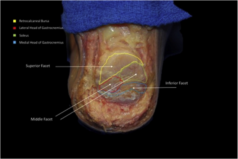
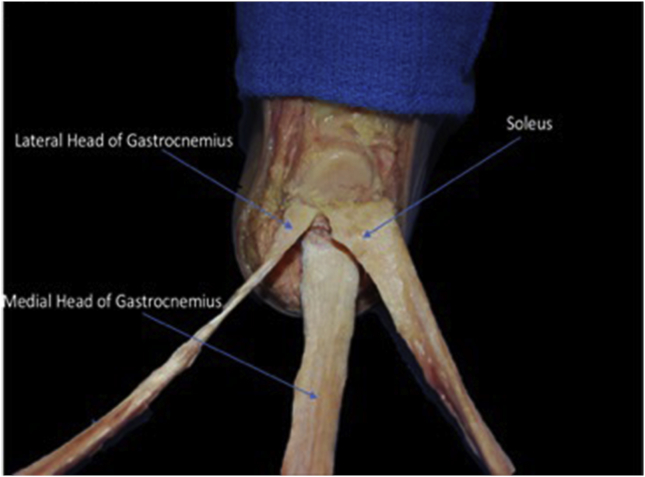
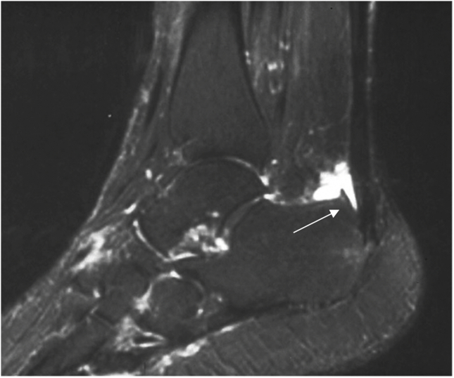
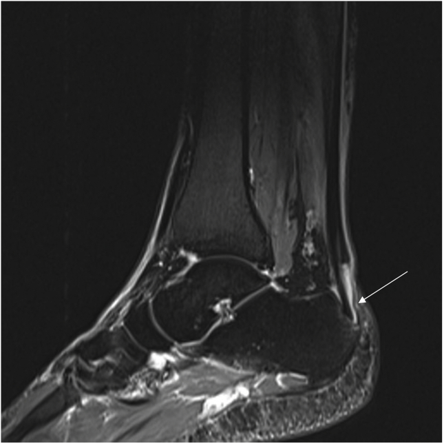
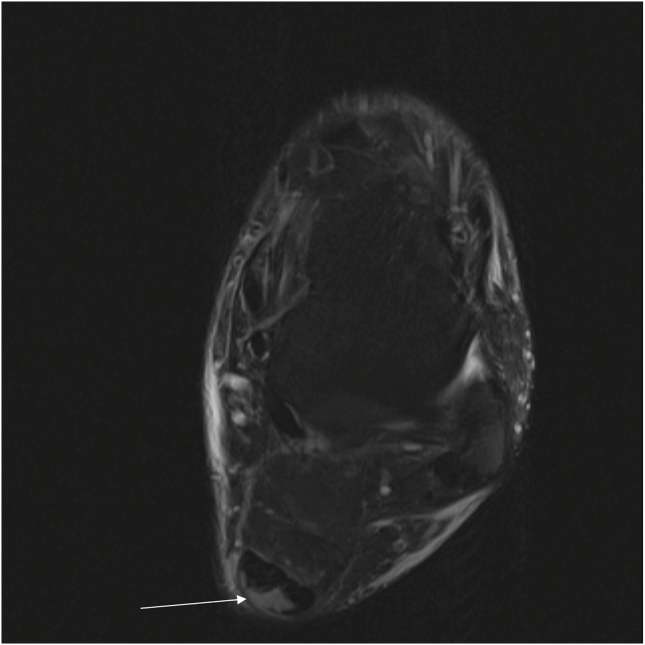
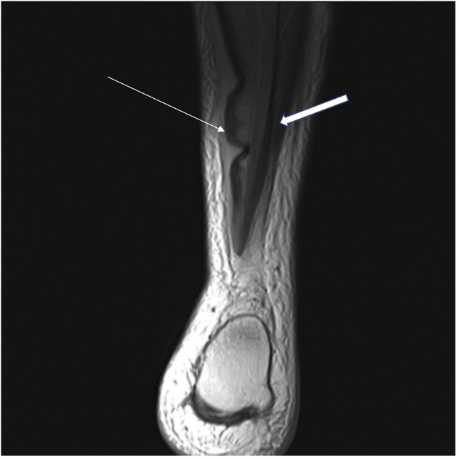
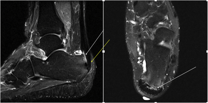
The Achilles tendon is the largest, and most commonly torn tendon in the body. The Achilles is usually torn at a region of relative hypo-vascularity proximal to its insertion. However, partial thickness tears and other pathologies often occur at its insertion on the calcaneus. Anatomically, the insertion is a confluence of the gastrocnemius and soleus muscles that fuse to form a myotendinous unit on the posterosuperior aspect of the calcaneus. This review aims to reveal the insertional footprint as individual fascicular components attaching to facets of calcaneal tuberosity. Understanding this anatomy is essential for interpreting tear patterns and surgical implications.
Keywords: Achilles complex; Achilles insertion; Achilles rupture; Achilles tear; Achilles tendon; Calcaneal tuberosity.
© 2019 Professor P K Surendran Memorial Education Foundation. Published by Elsevier B.V. All rights reserved.
Figures

References
-
- Ballal M.S., Walker C.R., Molloy A.P. The anatomical footprint of the Achilles tendon: a cadaveric study. Bone Joint Lett J. 2014;96-B(10):1344–1348. - PubMed
-
- Anson B.J., McVay C.B. Surgical Anatomy. fifth ed. WB Saunders Company; Philadelphia: 1971. pp. 1186–1189.
-
- Wood Jones F. Bailliere, Tindall and Cox; London: 1944. Structure and Function as Seen in the Foot; pp. 124–133.
-
- Szaro P., Witkowski G., Smigielski R., Krajewski P., Ciszek B. Fascicles of the adult human Achilles tendon: an anatomical study. Ann Anat. 2009;191:586–593. - PubMed
Publication types
LinkOut - more resources
Full Text Sources
