. 2019 Dec 19:19:342.
doi: 10.1186/s12935-019-1061-1. eCollection 2019.

Sulforaphene induces apoptosis and inhibits the invasion of esophageal cancer cells through MSK2/CREB/Bcl-2 and cadherin pathway in vivo and in vitro

Affiliations

Sulforaphene induces apoptosis and inhibits the invasion of esophageal cancer cells through MSK2/CREB/Bcl-2 and cadherin pathway in vivo and in vitro

Chengjuan Zhang et al. Cancer Cell Int. .

Abstract

Background: As a novel type of isothiocyanate derived from radish seeds from cruciferous vegetables, sulforaphene (SFE, 4-methylsufinyl-3-butenyl isothiocyanate) has various important biological effects, such as anti-oxidative and anti-bacterial effects. Recently, sulforaphene has attracted increasing attention for its anti-tumor effects and its ability to suppress the development of multiple tumors through different regulatory mechanisms. However, it has not yet been widely investigated for the treatment of esophageal cancer.

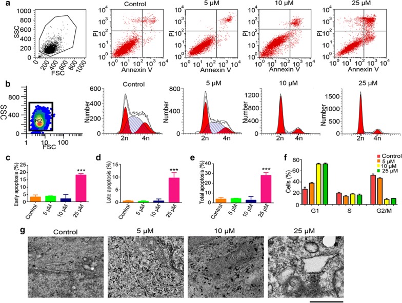
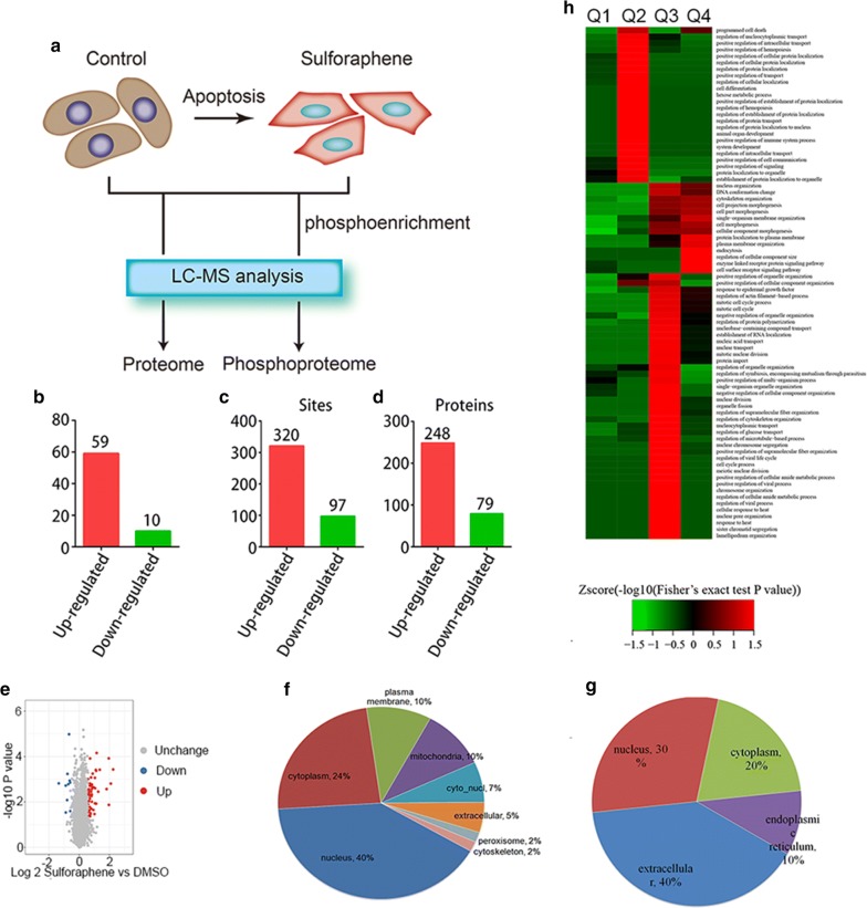
Methods: We observed an increased apoptosis in esophageal cancer cells on sulforaphene treatment through flow cytometry (FCM) analysis and transmission electron microscopy (TEM). Through mass spectrometry (MS) analysis, we further detected global changes in the proteomes and phosphoproteomes of esophageal cancer cells on sulforaphene treatment. The molecular mechanism of sulforaphene was verified by western blot,the effect and mechanism of SFE on esophageal cancer was further verified by patient-derived xenograft mouse model.

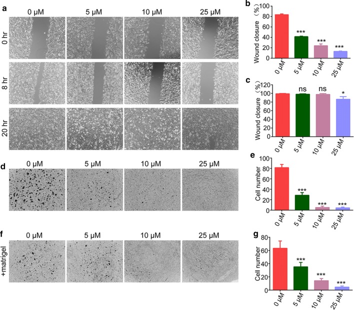
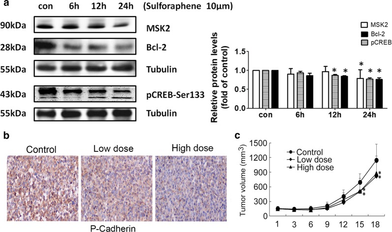
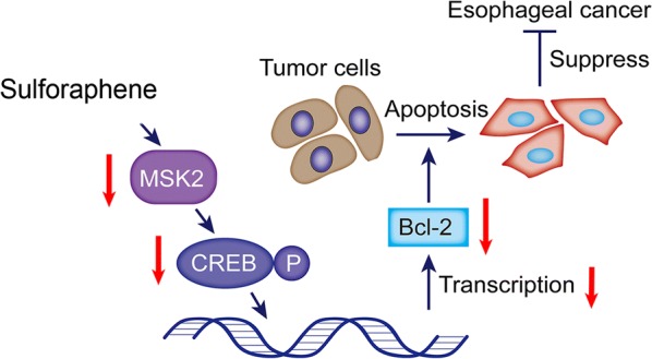
Results: We identified multiple cellular processes that were changed after sulforaphene treatment by proteomics. We found that sulforaphene could repress the phosphorylation of CREB through MSK2, leading to suppression of Bcl-2 and further promoted cell apoptosis. Additionally, we confirmed that sulforaphene induces tumor cell apoptosis in mice. Interestingly, we also observed the obvious inhibition of cell migration and invasion caused by sulforaphene treatment by inhibiting the expression of cadherin, indicating the complex effects of sulforaphene on the development of esophageal cancer.

Conclusions: Our data demonstrated that sulforaphene induced cell apoptosis and inhibits the invasion of esophageal cancer through a mechanism involving the inhibition of the MSK2-CREB-Bcl2 and cadherin pathway. Sulforaphene could therefore serve as a promising anti-tumor drug for the treatment of esophageal cancer.

Keywords: Apoptosis; Esophageal cancer; Invasion; MSK2; Sulforaphene.


Conflict of interest statement

Competing interestsThe authors declare that they have no competing interests.

Figures

Fig. 1
Fig. 1
Sulforaphene induces esophageal cancer cell apoptosis. a The flow cytometry profile represents the results of annexin V-FITC and PI staining and shows apoptosis in Eca109 cells. b The flow cytometry profile represents the results of PI staining and shows the cell cycle of Eca109 cells. c Comparison of early apoptosis rates in Eca109 cells with control and sulforaphene treatment. d The difference in the late apoptosis rates in Eca109 cells with control and sulforaphene treatment. e Comparison of the total apoptosis rates in Eca109 cells with control and sulforaphene treatment. f Comparison of the number of cells in the G1, S, and G2/M phases in Eca109 cells with control and sulforaphene treatment. g Representative TEM photographs showing cellular surfaces with short microvilli, an amorphous and granular glycocalyx, goblet cells full of mucigen granules and a large number of interdigitations. Data are expressed as the mean SD. * Indicates P < 0.05, **P < 0.01 and ***P < 0.001
Fig. 2
Fig. 2
Sulforaphene inhibits the migration and invasion of esophageal cancer cells in vitro. ac Wound healing assays were performed using Eca109 cells treated with DMSO or sulforaphene (5, 10, 25 μM), and the percentage of cell migration was measured after 8 or 20 h. d, e Transwell assays using DMSO- or sulforaphene-treated (5, 10, or 25 μM) Eca109 cells were performed, and the extent of Transwell migration was quantified by counting the cells. f, g Matrigel-based Transwell assays using DMSO- or sulforaphene-treated (5, 10, 25 μM) Eca109 cells were performed, and the extent of Transwell migration was quantified by cell counting. The results are presented as the mean ± SD
Fig. 3
Fig. 3
Differentially expressed proteins and phosphorylated proteins induced by sulforaphene treatment. a The flow chart of the experiment. Global protein expression and phosphorylation levels in the control and sulforaphene treatment groups were detected by mass spectrometry. b Significantly upregulated and downregulated proteins upon sulforaphene treatment were identified through mass spectrometry assays. c The number of significantly upregulated and downregulated phosphorylated protein sites upon sulforaphene treatment. d The number of significantly upregulated and downregulated phosphorylated proteins upon sulforaphene treatment. e Volcano plots of differentially expressed proteins. f The cellular distribution of significantly upregulated proteins. g The cellular distribution of significantly downregulated proteins. h GO analysis heat map of differentially expressed proteins
Fig. 4
Fig. 4
Sulforaphene induce tumor apoptosis through MSK2–CREB–Bcl-2 pathway. a Immunoblot analysis of the indicated MSK2, CREB and Bcl-2 expression levels in Eca109 cells with quantification data (n = 3). b IHC assays showed the expression levels of Bcl-2, and p-cadherin in both control and sulforaphene-treated tumor tissues isolated from mice. Data are expressed as the mean ± SD. * Indicates P < 0.05. c Comparisons of tumor volumes in representative mice in the control, low, and high sulforaphene-treated groups
Fig. 5
Fig. 5
Proposed molecular model of the role of sulforaphene in the esophageal cancer cell. Sulforaphene inhibited the MSK2, and CREB, to restrain the expression of the apoptosis protein Bcl-2, thereby facilitating the apoptosis of esophageal cancer cells and inhibiting cancer development

References

    1. Mizuta H, et al. Predictive factors for esophageal stenosis after endoscopic submucosal dissection for superficial esophageal cancer. Dis Esophagus. 2009;22(7):626–631. - PubMed
    1. Di Pardo BJ, et al. The global burden of esophageal cancer: a disability-adjusted life-year approach. World J Surg. 2016;40(2):395–401. - PubMed
    1. Lu HB. MicroRNA-556-3p promotes the progression of esophageal cancer via targeting DAB2IP. Eur Rev Med Pharmacol Sci. 2018;22(20):6816–6823. - PubMed
    1. Zhang SW, et al. Mortality and survival analysis of esophageal cancer in China. Zhonghua Zhong Liu Za Zhi. 2016;38(9):709–715. - PubMed
    1. He Y, et al. Incidence and mortality rate of esophageal cancer has decreased during past 40 years in Hebei Province, China. Chin J Cancer Res. 2015;27(6):562–571. - PMC - PubMed