. 2019 Dec 19:19:346.
doi: 10.1186/s12935-019-1059-8. eCollection 2019.

AGI-134: a fully synthetic α-Gal glycolipid that converts tumors into in situ autologous vaccines, induces anti-tumor immunity and is synergistic with an anti-PD-1 antibody in mouse melanoma models

Affiliations

AGI-134: a fully synthetic α-Gal glycolipid that converts tumors into in situ autologous vaccines, induces anti-tumor immunity and is synergistic with an anti-PD-1 antibody in mouse melanoma models

Stephen M Shaw et al. Cancer Cell Int. .

Abstract

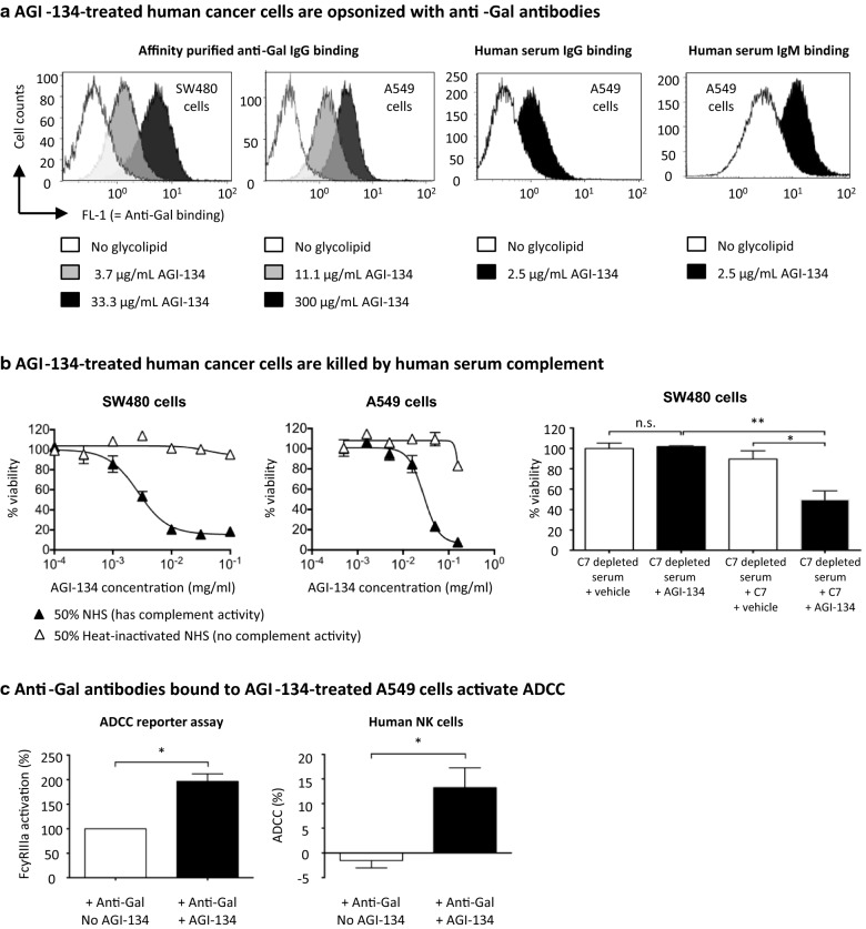
Background: Treatments that generate T cell-mediated immunity to a patient's unique neoantigens are the current holy grail of cancer immunotherapy. In particular, treatments that do not require cumbersome and individualized ex vivo processing or manufacturing processes are especially sought after. Here we report that AGI-134, a glycolipid-like small molecule, can be used for coating tumor cells with the xenoantigen Galα1-3Galβ1-4GlcNAc (α-Gal) in situ leading to opsonization with pre-existing natural anti-α-Gal antibodies (in short anti-Gal), which triggers immune cascades resulting in T cell mediated anti-tumor immunity.

Methods: Various immunological effects of coating tumor cells with α-Gal via AGI-134 in vitro were measured by flow cytometry: (1) opsonization with anti-Gal and complement, (2) antibody-dependent cell-mediated cytotoxicity (ADCC) by NK cells, and (3) phagocytosis and antigen cross-presentation by antigen presenting cells (APCs). A viability kit was used to test AGI-134 mediated complement dependent cytotoxicity (CDC) in cancer cells. The anti-tumoral activity of AGI-134 alone or in combination with an anti-programmed death-1 (anti-PD-1) antibody was tested in melanoma models in anti-Gal expressing galactosyltransferase knockout (α1,3GT-/-) mice. CDC and phagocytosis data were analyzed by one-way ANOVA, ADCC results by paired t-test, distal tumor growth by Mantel-Cox test, C5a data by Mann-Whitney test, and single tumor regression by repeated measures analysis.

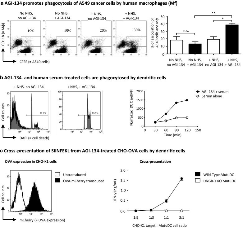
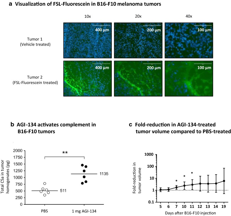
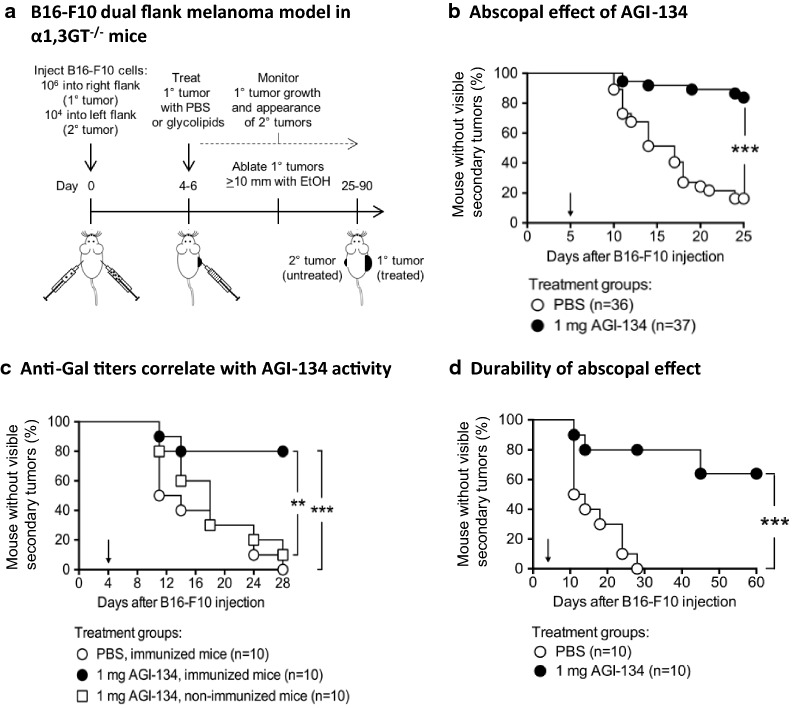
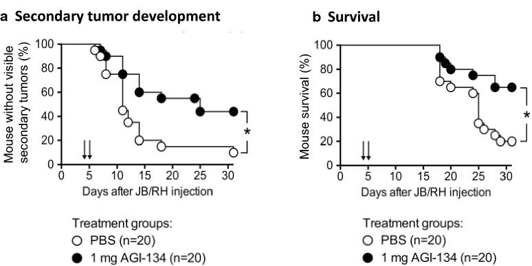
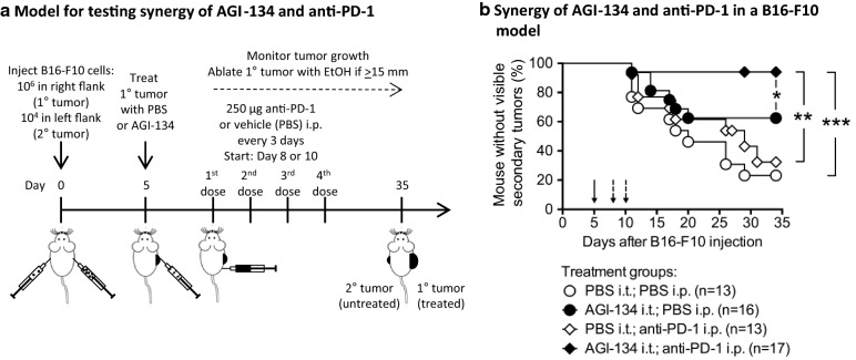
Results: In vitro, α-Gal labelling of tumor cells via AGI-134 incorporation into the cell membrane leads to anti-Gal binding and complement activation. Through the effects of complement and ADCC, tumor cells are lysed and tumor antigen uptake by APCs increased. Antigen associated with lysed cells is cross-presented by CD8α+ dendritic cells leading to activation of antigen-specific CD8+ T cells. In B16-F10 or JB/RH melanoma models in α1,3GT-/- mice, intratumoral AGI-134 administration leads to primary tumor regression and has a robust abscopal effect, i.e., it protects from the development of distal, uninjected lesions. Combinations of AGI-134 and anti-PD-1 antibody shows a synergistic benefit in protection from secondary tumor growth.

Conclusions: We have identified AGI-134 as an immunotherapeutic drug candidate, which could be an excellent combination partner for anti-PD-1 therapy, by facilitating tumor antigen processing and increasing the repertoire of tumor-specific T cells prior to anti-PD-1 treatment.

Keywords: AGI-134; Abscopal effect; Cancer vaccine; Checkpoint inhibition; Immunotherapy; Intratumoral injection; Melanoma; alpha-Gal; anti-Gal; anti-PD-1.


Conflict of interest statement

Competing interestsAll of the authors are or were employees, have or had executive roles and/or are consultants to, and may have stocks or shares of Agalimmune Ltd. or BioLineRx, and may hold patents related to the described work. Several of the authors are or were involved in efforts to develop AGI-134 as cancer immunotherapy. AGI-134 is currently in Phase 1/2a clinical trials in the United Kingdom and Israel with BioLineRx as sponsor.

Figures

Fig. 1
Fig. 1
Anti-Gal binds to AGI-134-treated human cancer cells and activates CDC and ADCC. a Human SW480 and A549 cancer cells were treated with PBS (open histograms) or the indicated concentrations of AGI-134 (grey and black histograms). The cells were then incubated with affinity purified human anti-Gal IgG or 25% heat-inactivated human serum. Anti-Gal antibody binding was detected with fluorescently-labeled secondary antibodies and samples analyzed by flow cytometry. Representative histogram overlays from two to three independently conducted experiments for each data set are shown. b SW480 and A549 cells were treated with half-log dilutions of AGI-134 and incubated with 50% normal (NHS) or heat-inactivated (iNHS) human serum. In some experiments, SW480 cells were exposed to C7 depleted serum ± 70 µg/mL C7. Cell viability was determined using a luminescence-based cell viability assay and data normalized and expressed as percentage viability. Representative data from 3 independent experiments are shown, with mean values ± SD. c A549 cells were treated with PBS or 0.5 mg/mL AGI-134 and then co-cultured with Promega’s ADCC reporter bioassay effector cells in a 25:1 effector:target cell ratio, in the presence or absence of 30 µg/mL affinity purified human anti-Gal IgG for 6 h. Induction of ADCC over no anti-Gal antibody controls was determined by addition of Bio-Glo Luciferase reagent to quantify reporter gene expression downstream of FcγRIIIa. For assessment of target cell killing by NK cells, CHO-K1 cells were treated with PBS or 1 mg/mL AGI-134 and pre-incubated with 30 µg/mL affinity purified human anti-Gal IgG, before co-culture with IL-2-activated human NK cells. After 4–6 h of co-culture the percentage of dead CHO-K1 cells was determined by incorporation of the viability dye 7-AAD into the target cells. Data shown is the mean + SEM for three (reporter bioassay) or six (cell killing assay) independent experiments
Fig. 2
Fig. 2
AGI-134-treated cells are phagocytosed by antigen-presenting cells and antigen cross-presented. a CFSE-labeled A549 cells were treated with PBS or 500 μg/mL AGI-134 and then incubated with or without normal human serum (NHS) to opsonize them with anti-Gal and complement. Subsequently, human macrophages were added at a A549 to macrophage ratio of 3:1. Subsequently, the co-cultures were stained with an anti-CD11 antibody and analyzed by flow cytometry. CFSE (for A549 cells) vs. CD11b (for macrophages) dot plots are shown for the various conditions. Double-positive events were assumed to be macrophages with associated (adherent or phagocytosed) A549 cells. In the bar graphs, the results of three independent experiments, specifically the average percentages of double positive events + SD are shown (*p < 0.05; **p < 0.005; ns, not significant; one-way ANOVA). b CHO-K1 cells were treated with 1 mg/ml AGI-134 and then with or without 50% NHS. Cell killing was determined by DAPI-staining of a cell aliquot. The range gates in the histogram plots quantified dead cells. The remaining CHO-K1 cells were stained with CellVue Claret dye and incubated with GFP-expressing MutuDC cells at 1:1 ratios. Samples were removed from the co-culture after 30–120 min, and analyzed by flow cytometry. The CellVue Claret dye geometric mean fluorescence intensities (gMFIs) were normalized as described in the methods and then plotted against time. c CHO-K1 cells were transduced to express OVA tagged with the fluorophore mCherry. The histogram shows an overlay for the mCherry signal for CHO-K1 parental cells (open curve) and CHO-OVA cells (closed curve). After treatment with vehicle or 1 mg/ml AGI-134, the CHO-OVA cells were incubated with 50% NHS before co-culture with wild-type or DNGR-1 KO MutuDCs at the indicated range of dead CHO-OVA:MutuDC cell ratios. After 4 h, OT-1 CD8+ T cells were added to the co-culture and incubated overnight. OT-1 T cell activation was quantified by IFN-γ ELISA of the co-culture supernatants
Fig. 3
Fig. 3
Primary tumor treatment with AGI-134 causes tumor regression, activates complement and FSL distribution in tumors. a Glycolipid detection in B16-F10 tumors: FSL-Fluorescein was used as surrogate molecule for AGI-134 visualization in tumors. 1 × 106 B16-F10 cells were grafted onto immunized α1,3GT−/− mice on both flanks. Five days later, the two tumors on each mouse were treated with 100 μL of 1 mg/mL FSL-fluorescein on one flank and with 100 μL PBS on the other flank. The following day, the tumors were excised and frozen in OCT compound. The tumors were sectioned and labeled with DAPI. Pictures in the GFP and DAPI channels for FSL and tumor cell nucleus DNA visualization were taken using ×4–×40 objectives (×10 example pictures are shown). The pictures show representative data of DAPI and GFP channel picture overlays for a vehicle- and a fluorescein-lipid treated tumor from the same mouse. b In complement activation experiments, B16-F10 tumors were treated by intratumoral injection of vehicle (PBS) or 1 mg AGI-134 on Day 6 post B16-F10 cell grafting. 2.5 h after treatment, tumors were excised, homogenized and the complement factor C5a measured by ELISA. Each symbol represents the total C5a in the tumor homogenate of each mouse, median C5a values are indicated by the bars. Differences between the PBS vs. AGI-134 treatment groups were assessed by Mann–Whitney test (**p < 0.003). c In primary tumor regression experiments, back transformed least square geometric means for PBS and AGI-134 treatments across the timepoints were calculated and fold-reduction in geometric means ± 95% CI plotted, (*p < 0.05, n = 13)
Fig. 4
Fig. 4
AGI-134 treatment of primary tumors produces an abscopal effect protecting mice from contralateral tumor development. a Schematic of abscopal B16-F10 melanoma model in anti-Gal-expressing α1,3GT−/− mice. To monitor the abscopal effect of AGI-134, primary B16-F10 lesions were treated once by intratumoral injection of PBS or 1 mg AGI-134 and the development of contralateral lesions monitored. The percentages of mice without visible/palpable contralateral tumors are plotted in the graphs. The solid arrows indicate the day of AGI-134 or mock treatment (Day 4–6). b The pooled data from four independent experiments where the abscopal effect in B16-F10 tumors was monitored over 25 days are summarized. c B16-F10 tumors in immunized (anti-Gal positive) or non-immunized (anti-Gal negative) α1,3GT−/− mice were treated i.t. with vehicle, or 1 mg AGI-134. d Representative data from two experiments where the abscopal effect of AGI-134 in B16-F10 tumors was monitored over 60–90 days is shown. Statistical differences between treatment groups in each plot were analyzed by Mantel–Cox test (**p < 0.005; ***p < 0.0005)
Fig. 5
Fig. 5
AGI-134 protects mice from secondary tumor development and improves survival in a JB/RH melanoma model. Anti-Gal-expressing α1,3GT−/− mice were grafted with 5 × 105 JB/RH cells to create a 1° tumor on one flank and 2 × 104 JB/RH cells on the contralateral flank. 4–5 days after grafting, the 1° tumors were treated once with 1 mg AGI-134 or PBS and contralateral tumor development (a) and mouse survival (b) monitored. Pooled data from three independent experiments are shown. Statistical differences between treatment groups were analyzed by Mantel-Cox test (*p < 0.05). The solid arrows indicate the day of AGI-134 or mock treatment (Day 4 or 5)
Fig. 6
Fig. 6
AGI-134 synergizes with an anti-PD-1 antibody. a Schematic for testing efficacy of AGI-134 in combination with RMP1-14, an anti-PD-1 antibody. b On Day 5 after B16-F10 cell grafting, mice were treated i.t. with single 100 or 250 µg doses of AGI-134 or vehicle, and then intraperitoneally with four 250-μg doses of RMP1-14 or vehicle in 3–4-day intervals starting on Day 8 (experiment #1) or Day 10 (experiment #2) post B16-F10 cell grafting. For the graph, the data from two independent experiments were combined and plotted. The data show the percentage of mice free from secondary tumors over time. The treatment groups were statistically compared by Mantel–Cox test (*p < 0.05; **p < 0.005; ***p < 0.0005). Solid arrows indicate the time of i.t. AGI-134 or vehicle treatment; dashed arrows show the start of i.p. RMP1-14 treatment

Similar articles

Cited by

References

    1. Balar AV, Weber JS. PD-1 and PD-L1 antibodies in cancer: current status and future directions. Cancer Immunol Immunother. 2017;66(5):551–564. doi: 10.1007/s00262-017-1954-6. - DOI - PMC - PubMed
    1. Sharma P, Allison JP. The future of immune checkpoint therapy. Science. 2015;348(6230):56–61. doi: 10.1126/science.aaa8172. - DOI - PubMed
    1. Sathyanarayanan V, Neelapu SS. Cancer immunotherapy: strategies for personalization and combinatorial approaches. Mol Oncol. 2015;9(10):2043–2053. doi: 10.1016/j.molonc.2015.10.009. - DOI - PMC - PubMed
    1. Bommareddy PK, Silk AW, Kaufman HL. Intratumoral approaches for the treatment of melanoma. Cancer J. 2017;23(1):40–47. doi: 10.1097/PPO.0000000000000234. - DOI - PubMed
    1. Galili U, et al. Man, apes, and Old World monkeys differ from other mammals in the expression of alpha-galactosyl epitopes on nucleated cells. J Biol Chem. 1988;263(33):17755–17762. - PubMed