

A case of solitary rectal diverticulum presenting with a large retrorectal abscess
- PMID: 31890199
- PMCID: PMC6926126
- DOI: 10.1016/j.amsu.2019.11.015
A case of solitary rectal diverticulum presenting with a large retrorectal abscess
Abstract
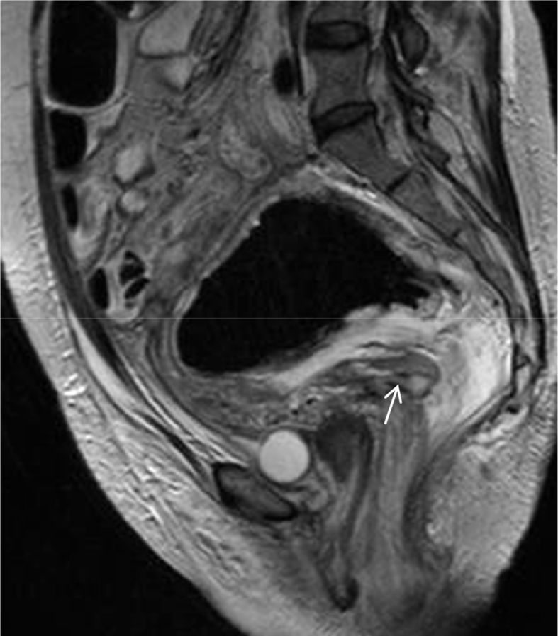
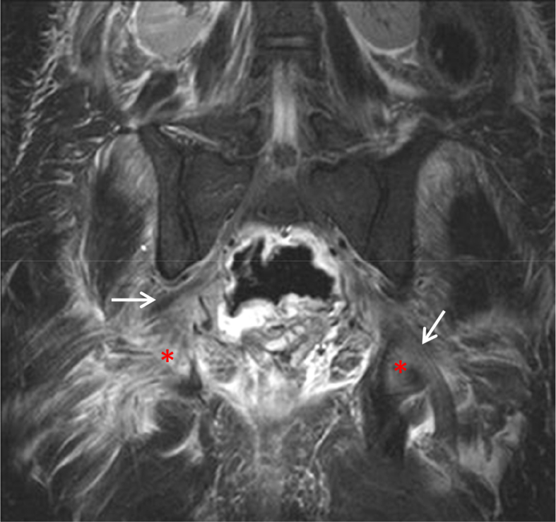
Colonic diverticular disease is a common condition, affecting 50% of the population aged above 80. In contrast, rectal diverticular disease is a rare condition with very few cases reported, while symptomatic rectal diverticular disease is even rarer. We present a case of a symptomatic large rectal diverticulum presenting with a retrorectal abscess. A 49-year-old Caucasian female was brought to the emergency department complaining of abdominal pain and weakness in the lower limbs. She was found to have obstructive uropathy and unilateral sciatic neuropathy. She rapidly developed acute abdomen and emergency laparotomy revealed a giant purulent rectal diverticulum. The patient underwent exploratory laparotomy and a loop colostomy was made to decompress the colon.
Keywords: Abscess; Complications; Diverticulitis; Rectal diverticulum.
© 2019 The Authors.
Conflict of interest statement
The Authors declare that have no Conflict of Interest.
Figures


References
-
- Alabiso M.E., Grassi R., Fioroni C., Marano I. Iatrogenic rectal diverticulum in patients treated with transanal stapled techniques. Radiol. Med. 2008;113:887–894. - PubMed
-
- Piercy K.T., Timaran C., Akin H. Rectal diverticula: report of a case and review of the literature. Dis. Colon Rectum. 2002;45:1116–1117. - PubMed
Publication types
LinkOut - more resources
Full Text Sources
