

Wide-field angiography in retinal vein occlusions
- PMID: 31890282
- PMCID: PMC6907103
- DOI: 10.1186/s40942-019-0163-1
Wide-field angiography in retinal vein occlusions
Abstract
Background: Retinal vein occlusion (RVO) is the second most common retinal vascular disease after diabetic retinopathy. It can result in significant visual loss from complications like macula edema, retinal and iris neovascularization, and vitreous hemorrhage. Recently, ultra-widefield imaging (UWF) has been developed for posterior pole visualization and has shown to be useful in the evaluation and treatment of RVO.
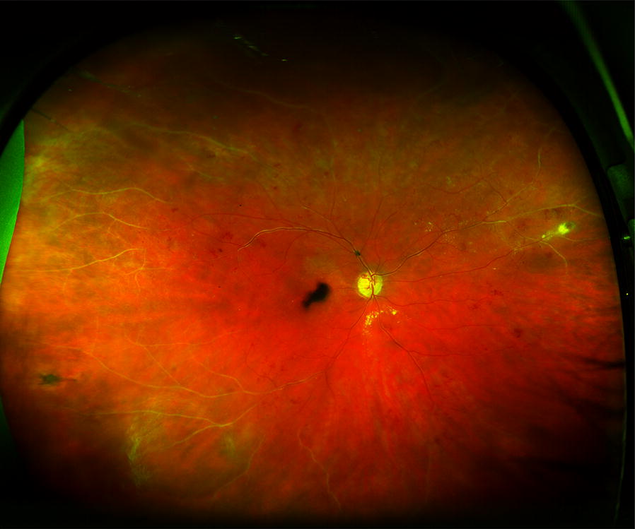
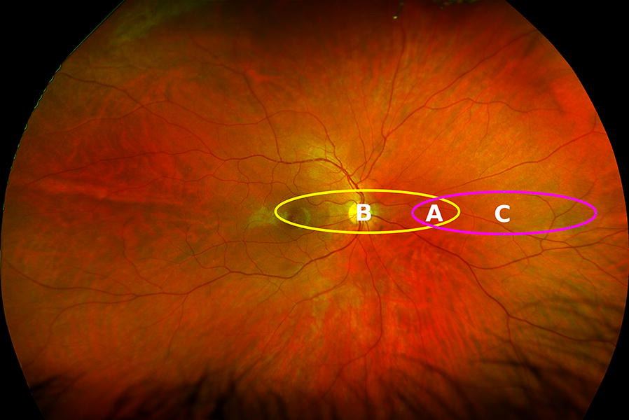
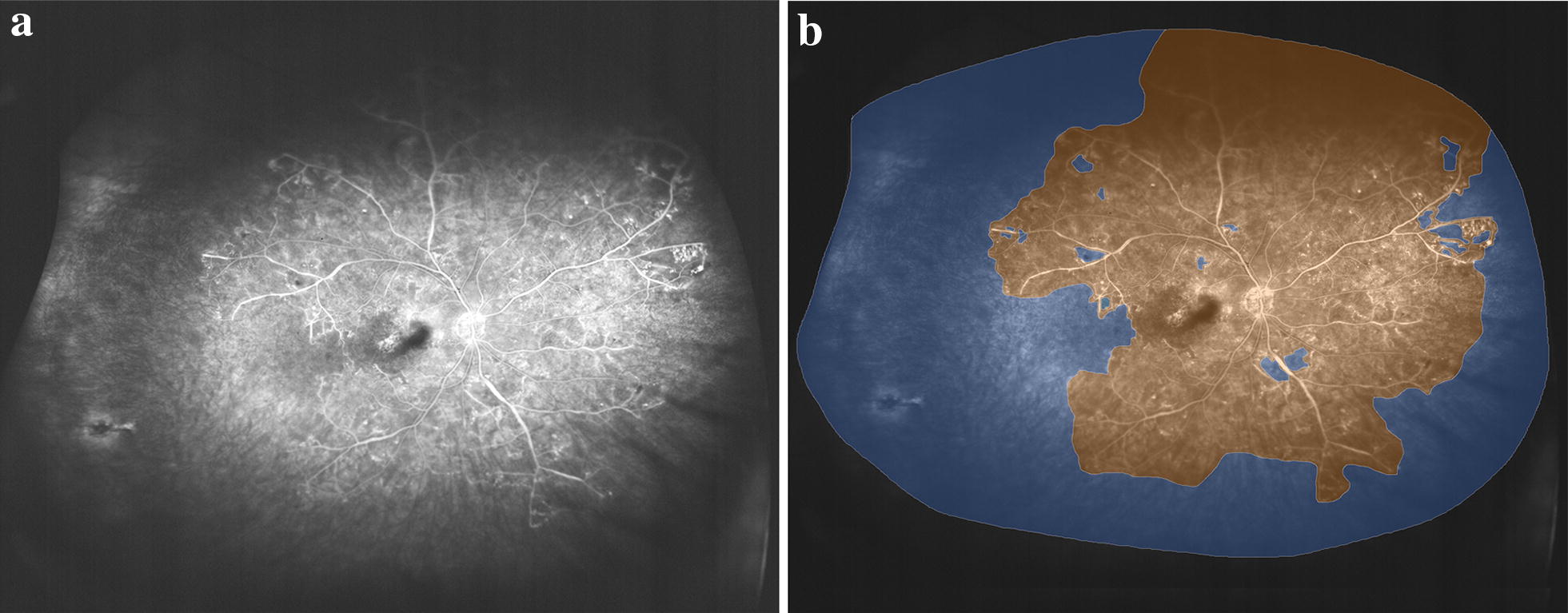
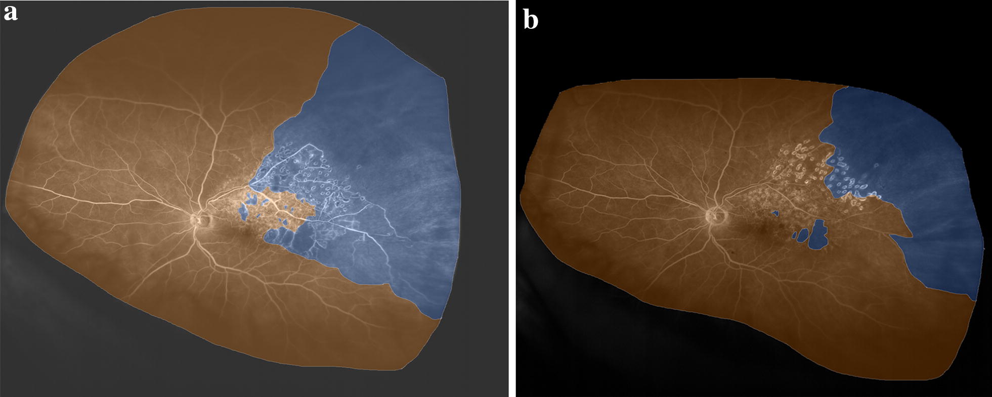
Main text: Ultra-widefield imaging (UWF) imaging allows for visualization of the retina up to an angle of 200°. This is especially important in detecting peripheral retinal pathologies, especially in retinal conditions such as RVO, where the disease process affects the peripheral as well as central retina. In particular, retinal non-perfusion in RVO is a risk factor for neovascularization. Various techniques, such as ischemic index and stereographic projection, have been described to assess areas of ischemia on UWF images. Retinal non-perfusion has an impact on disease complications, such as macular edema, and retinal and iris neovascularization. Retinal non-perfusion also has implications on disease response, including visual acuity, reduction in retinal edema and treatment burden.
Conclusion: Ultra-widefield imaging (UWF) imaging plays an important role in the assessment and management of RVO, especially in measuring retinal non-perfusion in the peripheries.
Keywords: Fluorescein angiography; Ischemic index; Macular edema; Retinal vein occlusion; Ultrawidefield imaging.
© The Author(s) 2019.
Conflict of interest statement
CST—Research Support from National Medical Research Council Transition Award (NMRC/TA/0039/2015). Conference support from Bayer, Heidelberg Engineering and Novartis. KZL—none. SRS—Consultant for Allegan, Genentech, Roche, Novartis, Iconic, Thrombogenics, Centervue, Heidelberg, Optos and Carl Zeiss Meditec. Research Support from Allergan, Genentech, Optos, and Carl Zeiss Meditec.
Figures
References
-
- Rogers SL, McIntosh RL, Lim L, Mitchell P, Cheung N, Kowalski JW, Nguyen HP, Wang JJ, Wong TY. Natural history of branch retinal vein occlusion: an evidence-based systematic review. Ophthalmology. 2010;117(1094–1101):e1095. - PubMed
Publication types
LinkOut - more resources
Full Text Sources
