Review
. 2019 Dec 12;5(Suppl 1):49.
doi: 10.1186/s40942-019-0196-5. eCollection 2019.

Widefield imaging of retinal and choroidal tumors

Affiliations
Review

Widefield imaging of retinal and choroidal tumors

Natalia F Callaway et al. Int J Retina Vitreous. .

Abstract

Background: Wide-field imaging plays an increasingly important role in ocular oncology clinics. The purpose of this review is to describe the commonly used wide-field imaging devices and review conditions seen in ocular oncology clinic that underwent wide-field imaging as part of the multimodal evaluation.

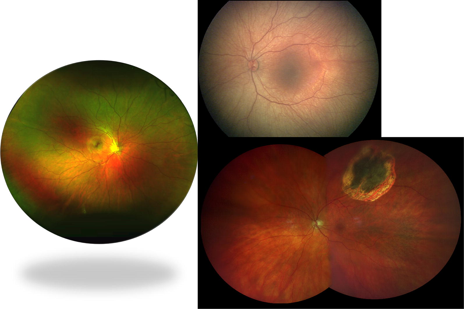
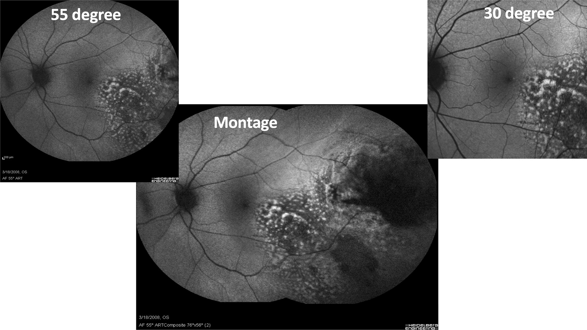
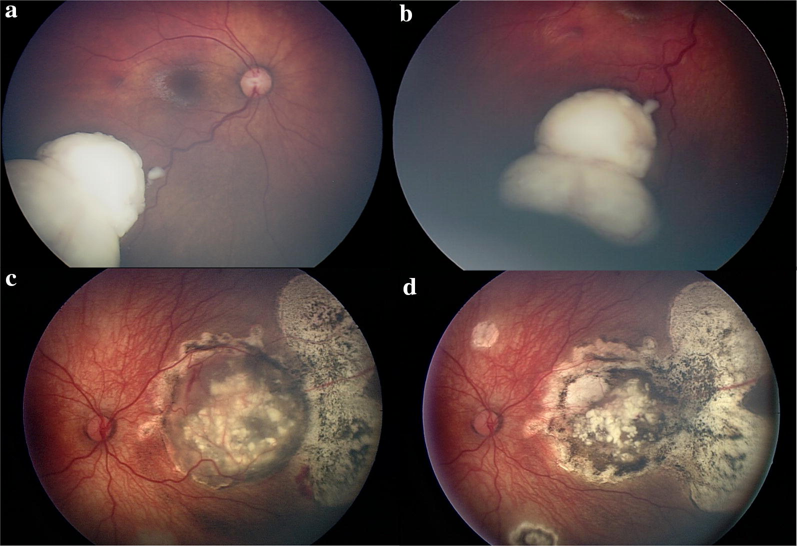
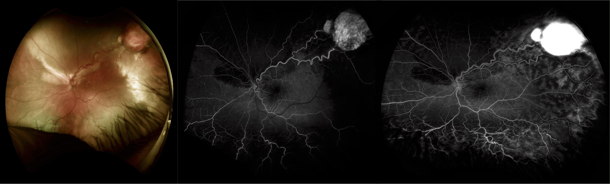
Summary of review: Wide-field or wide-angle imaging is defined as greater than 50° field of view. Modern devices can reach far beyond this reporting fields of view up to 267°, when utilizing montage features, with increasingly impressive resolution. Wide-field imaging modalities include fundus photography, fluorescein angiography (FA), fundus autofluorescence (FAF), indocyanine angiography (ICG), spectral domain optical coherence tomography (SD-OCT), and recently wide-field OCT Angiography (OCTA). These imaging modalities are increasingly prevalent in practice. The wide-field systems include laser, optical, and lens based systems that are contact or non-contact lens systems each with its own benefits and drawbacks. The purpose of this review is to discuss commonly used wide-field imaging modalities for retinal and choroidal tumors and demonstrate the use of various widefield imaging modalities in select ocular oncology cases.

Conclusions: Clinical examination remains the gold standard for the evaluation of choroidal and retinal tumors. Wide-field imaging plays an important role in ocular oncology for initial documentation, surgical planning, determining the relationship of the tumor to adjacent ocular structures, following tumor size after treatment, and monitoring for recurrence.

Keywords: Choroid; Clarus 500; Eye; Heidelberg Spectralis; Hemangioma; Melanoma; Metastasis; Nevus; Optos; Panoret-1000; Phoenix Icon; RetCam 3; Retina; Retinoblastoma; Tumor; Wide-field angiography; Wide-field imaging; confocal scanning laser ophthalmoscopy; wide-field photography.


Conflict of interest statement

Competing interestsNFC has no competing interests. PM is a consultant for Optos.

Figures

Fig. 1
Fig. 1
Field of view with various widefield imaging systems. Left: Optos camera (Optos PLC, Dunfermline, UK) offers up to 200° of horizontal field of view. Right Upper: 130° RetCam 3 (Clarity Medical Systems, Inc., Pleasanton, CA, USA). Right Lower: Clarus 500 (Carl Zeiss Meditech Inc., Dublin, CA)
Fig. 2
Fig. 2
Fundus autofluorescence of a choroidal melanoma with the Heidelberg Spectralis (Heidelberg, Engineering, Inc., Heidlberg, Germany) using a standard 30° image (upper right), an extended 55° image (upper left), and a montage image (center). The choroidal melanoma demonstrates increase hyperautofluorescense corresponding to clinical areas of orange pigment as well as RPE changes
Fig. 3
Fig. 3
Retinoblastoma imaging with RetCam 3 (Clarity Medical Systems, Inc., Pleasanton, CA, USA). a Retinoblastoma along the inferotemporal arcade with dilated feeding and draining retinal vessels. b Retinoblastoma anterior margin captured with scleral depression. c Large nasal retinoblastoma with calcifications and chorioretinal changes in the periphery. d Shows regression of the tumor following treatment
Fig. 4
Fig. 4
Optos wide-field fundus photo (left) and FA (middle and right images) of a temporal VHL associated retinal hemangioblastoma in the left eye. Wide-field imaging and FA play an important role as the lesions are often peripheral and demonstrate a classic FA pattern of rapid filling of the feeding retinal arteriole, later filling of the intralesional blood vessels, followed by drainage through he dilated venule Left Optos wide-field image. Middle: Early phase (31 s) at arteriolar filling demonstrating rapid dye. Right: Later phase (1:02 min) with rapid dye filling into the hemangioblastoma and leakage of the dye
Fig. 5
Fig. 5
Wyburn-Mason Racemose Hemangioma Optos Wide-field Fundus Imaging on the left and Fluorescein Angiography on the right. Fundus photo demonstrates a large racemose hemangioma with a temporal sclerotic vessel. The fluorescein angiography reveals temporal non-perfusion. These images were originally published in the Retina Image Bank® website. Authors: Sarina Amin and Neal Palejwala. Photographer: Sarah Ellano. Title: Wyburn-Mason Widefield Fundus Photography and Fluorescein Angiography. Retina Image Bank. April 29 2018. © the American Society of Retina Specialists. Images 28152 and 28153
Fig. 6
Fig. 6
Multimodal imaging in the diagnosis of a uveal melanoma. The patient presented with symptoms of vertical ribbons in his vision and was referred to oncology after fundus examination revealed an elevated lesion along the superotemporal arcade show in the standard photograph (a). OCT through the macula (b) demonstrates temporal subretinal fluid (arrow) corresponding to the subretinal fluid and associated pigmentary changes seen on the Optos wide-field images (c). Fundus autofluorescence of the macula (d) and wide-field with Optos (e) highlight the hyperautofluorescence of the subretinal fluid track.
Fig. 7
Fig. 7
multimodal imaging of uveal melanoma. a Standard fundus photography demonstrating a choroidal mass with overlying retinal pigmentary changes and small area of detachment and subretinal fluid. b Optos wide-field imaging fo the same mass capturing the peripheral margins of the tumor. c Optos fluorescein angiography demonstrates irregular filling of the mass without other lesions visible in the retina. d Wide-field fundus autofluorescence of the tumor reveals hypo autofluorescence of the mass with inferior hyperautofluorescence consistent with subretinal fluid on examination
Fig. 8
Fig. 8
Multimodal imaging of a uveal metastasis from lung adenocarcinoma. The patient presented with reduced vision in his right eye and clinical examination revealed a large, placoid-like mass at the superotemporal arcade with overlying pigmentary changes and associated fluid. a Optos wide-field imaging of the placoid lesion with overlying pigmentary changes. b OCT with en face image with pigmentary changes, irregular, low-lying choroidal mass and resulting undulations of the choroid and associated subretinal fluid. c Ultrasonography reveals the lesion is irregular. d After target treatment the lesion thickness decreases on ultrasonography and e the choroidal mass has resolved and the subretinal fluid absorbed
Fig. 9
Fig. 9
Radiation retinopathy following melanoma treatment. a Optos wide-field image of radiation retinopathy following melanoma treatment with brachytherapy. Superonasal to the disc is a flat chorioretinal scar with retinal hemorrhage and adjacent pigmentary changes. b Wide-field Optos fluorescein angiography reveals nonperfusion in the area of the scar with late leakage at the border with early development of radiation retinopathy. c Montage fundus photo demonstrating superonasal hemorrhage and radiation retinopathy

References

    1. Shields CL, Manalac J, Das C, Saktanasate J, Shields JA. Review of spectral domain enhanced depth imaging optical coherence tomography of tumors of the choroid. Indian J Ophthalmol. 2015;63:117–121. doi: 10.4103/0301-4738.154377. - DOI - PMC - PubMed
    1. McNabb RP, Grewal DS, Mehta R, Schuman SG, Izatt JA, Mahmoud TH, Jaffe GJ, Mruthyunjaya P, Kuo AN. Wide field of view swept-source optical coherence tomography for peripheral retinal disease. Br J Ophthalmol. 2016;100:1377–1382. doi: 10.1136/bjophthalmol-2015-307480. - DOI - PMC - PubMed
    1. Shields CL, Shields JA, Kiratli H, De Potter P, Cater JR. Risk factors for growth and metastasis of small choroidal melanocytic lesions. Trans Am Ophthalmol Soc. 1995;93:259–275. - PMC - PubMed
    1. Kim JW, Ngai LK, Sadda S, Murakami Y, Lee DK, Murphree AL. Retcam fluorescein angiography findings in eyes with advanced retinoblastoma. Br J Ophthalmol. 2014;98:1666–1671. doi: 10.1136/bjophthalmol-2014-305180. - DOI - PubMed
    1. Seider MI, Grewal DS, Mruthyunjaya P. Portable optical coherence tomography detection or confirmation of ophthalmoscopically invisible or indeterminate active retinoblastoma. Ophthalmic Surg Lasers Imaging Retina. 2016;47:965–968. doi: 10.3928/23258160-20161004-12. - DOI - PubMed