

Fresh Osteochondral Resurfacing of the Patellofemoral Joint
- PMID: 31890513
- PMCID: PMC6926379
- DOI: 10.1016/j.eats.2019.07.017
Fresh Osteochondral Resurfacing of the Patellofemoral Joint
Abstract
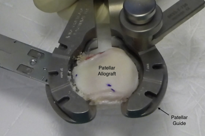
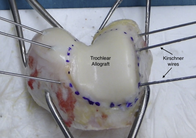
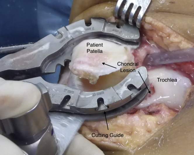
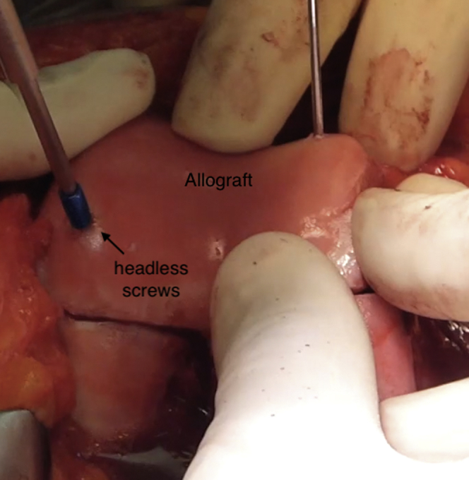
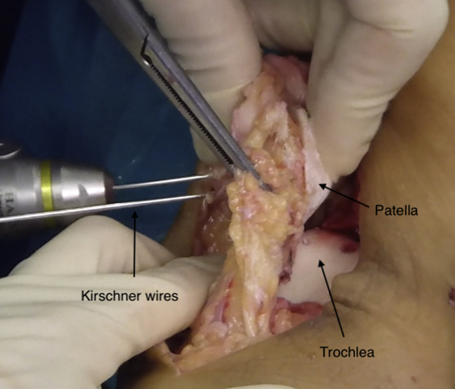
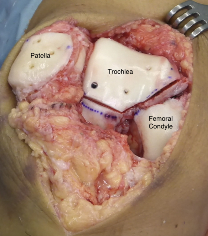
Large osteochondral lesions of the knee in young patients continue to be a challenge for orthopaedic surgeons and the focus of continual research. This is particularly true if the injury is a consequence of a dysplastic trochlea and involves both articular surfaces of the biomechanically complex patellofemoral joint. To obtain a healthy and congruent patellofemoral joint, the use of a bipolar fresh osteochondral allograft transplantation of the patella and trochlea is one of the few options to biologically treat these injuries. This would achieve a replacement of the entire articular surface of the patellofemoral joint with a high number of viable chondrocytes and respect the unique structural characteristics of the cartilage. The aim of this study was to obtain symptomatic and functional improvements while delaying the timing of prosthetic surgery. We present a reproducible although demanding surgical technique to perform a bipolar fresh osteochondral allograft transplantation of the patella and trochlea.
© 2019 by the Arthroscopy Association of North America. Published by Elsevier.
Figures

References
-
- Draper C., Bezier T., Gold G., Fredericson M., Fiene A., Beaupre A. Is cartilage thickness different in young subjects with and without patellofemoral pain? Osteoarthritis Cartilage. 2006;14:931–937. - PubMed
-
- Mason J.J., Leszko F., Johnson T., Komistek R.D. Patellofemoral joint forces. J Biomech. 2008;41:2337–2348. - PubMed
-
- Brophy R.H., Wojahn R.D., Lamplot J.D. Cartilage restoration techniques for the patellofemoral joint. J Am Acad Orthop Surg. 2017;25:321–329. - PubMed
LinkOut - more resources
Full Text Sources
