

Case Reports
doi: 10.1016/j.eucr.2019.101096.
eCollection 2020 Mar.
Seminoma metastasized to the prostate: A case report and literature review
Affiliations
- PMID: 31890599
- PMCID: PMC6928271
- DOI: 10.1016/j.eucr.2019.101096
Item in Clipboard
Case Reports
Seminoma metastasized to the prostate: A case report and literature review
Urol Case Rep.
.
Abstract
The testicular seminomas are germ-cell tumors which account for approximately 50% of all testicular tumors. Most primary testicular germ cell tumors metastasize through a lymphatic system in a predictable pattern with the retroperitoneal lymph nodes being the most common initial metastatic site. Hematological metastasis to the distant organs is less common, and except for pulmonary metastasis, changes the classification from good to intermediate prognosis. Metastasis of testicular seminoma to the prostate is an extremely rare entity with only five reported cases in the literature. In this report, we present a 63-year-old male with recurrent testicular seminoma presenting in prostate.
© 2019 The Authors.
Figures
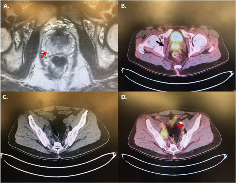
MRI of the pelvis (A) showed a 3 cm mass (red arrow) at the right basilar central zone of prostate. The mass was hyperintense in PET-CT scan involving the base of the prostate and seminal vesicle. (B, black arrow). Bilateral pelvic lymphadenopathies were noted in surveillance CT-scan (C) and they were hyperintense in PET-CT scan. (D, red arrowhead). (For interpretation of the references to colour in this figure legend, the reader is referred to the Web version of this article.)
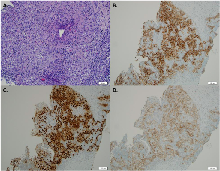
Tumor cells show pink to focally clear cytoplasm, vesicular nuclei and lymphocytic infiltration (A). Tumor cells are positive for PLAP (B), OCT3/4 (C) and CD117 (D), and negative for pancytokeratin (not shown). (For interpretation of the references to colour in this figure legend, the reader is referred to the Web version of this article.)
Similar articles
-
Testicular seminoma metastasis to the gastrointestinal tract and the necessity of surgery.J Gastrointest Cancer. 2012 Sep;43(3):499-501. doi: 10.1007/s12029-011-9274-0. J Gastrointest Cancer. 2012. PMID: 21519812
-
Metastatic Seminoma Presenting in Kidney and Cervical Lymph Nodes after a 25-Year Interval: A Case Report and Literature Review.Case Rep Oncol. 2023 Nov 29;16(1):1508-1517. doi: 10.1159/000535026. eCollection 2023 Jan-Dec. Case Rep Oncol. 2023. PMID: 38033414 Free PMC article.
-
[Positron emission tomography with [18 F]-2-fluoro-2-deoxy-D-glucose (18FDG-PET) in diagnosis of retroperitoneal lymph node metastases of testicular tumors].Urologe A. 1998 Nov;37(6):609-20. doi: 10.1007/s001200050223. Urologe A. 1998. PMID: 9887489 German.
-
Management of testicular seminoma advanced disease. Report on 14 cases and review of the literature.Arch Ital Urol Androl. 2002 Jun;74(2):81-5. Arch Ital Urol Androl. 2002. PMID: 12161942 Review.
-
[Treatment and prognosis of testicular seminoma].Gan To Kagaku Ryoho. 2000 Apr;27(4):516-21. Gan To Kagaku Ryoho. 2000. PMID: 10790992 Review. Japanese.
Cited by
-
A case of prostatic metastasis from non-seminomatous testicular cancer.IJU Case Rep. 2023 Jun 27;6(5):274-277. doi: 10.1002/iju5.12602. eCollection 2023 Sep. IJU Case Rep. 2023. PMID: 37667767 Free PMC article.
-
Robot-Assisted Laparoscopic Radical Prostatectomy for Prostatic Metastatic Recurrence from Testicular Cancer.Case Rep Urol. 2024 Jun 11;2024:1941414. doi: 10.1155/2024/1941414. eCollection 2024. Case Rep Urol. 2024. PMID: 38898921 Free PMC article.
-
Metastatic Seminoma with Positive Staining of Cytokeratin and MOC31: A Diagnostic Pitfall.Case Rep Pathol. 2021 Jun 29;2021:9992978. doi: 10.1155/2021/9992978. eCollection 2021. Case Rep Pathol. 2021. PMID: 34306787 Free PMC article.
References
-
- Bray F., Ferlay J., Soerjomataram I., Siegel R.L., Torre L.A., Jemal A. Global cancer statistics 2018: GLOBOCAN estimates of incidence and mortality worldwide for 36 cancers in 185 countries. CA A Cancer J Clin. 2018;68(6):394–424. - PubMed
-
- National Comprehensive Cancer Network Testicular cancer (version 1.2019. https://www.nccn.org/professionals/physician_gls/pdf/testicular.pdf
-
- Warde P., Specht L., Horwich A. Prognostic factors for relapse in stage I seminoma managed by surveillance: a pooled analysis. J Clin Oncol. 2002;20(22):4448–4452. - PubMed
Publication types
LinkOut - more resources
Full Text Sources
