Review
. 2019 Dec 27:7:39.
doi: 10.1186/s41038-019-0175-y. eCollection 2019.

Diagnosis and Treatment of Keloids and Hypertrophic Scars-Japan Scar Workshop Consensus Document 2018

Affiliations
Review

Diagnosis and Treatment of Keloids and Hypertrophic Scars-Japan Scar Workshop Consensus Document 2018

Rei Ogawa et al. Burns Trauma. .

Abstract

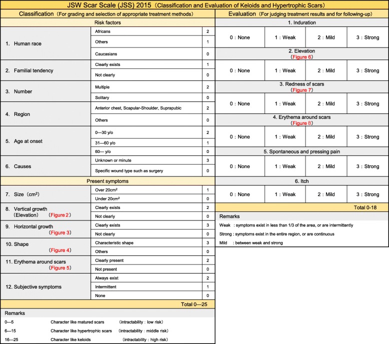
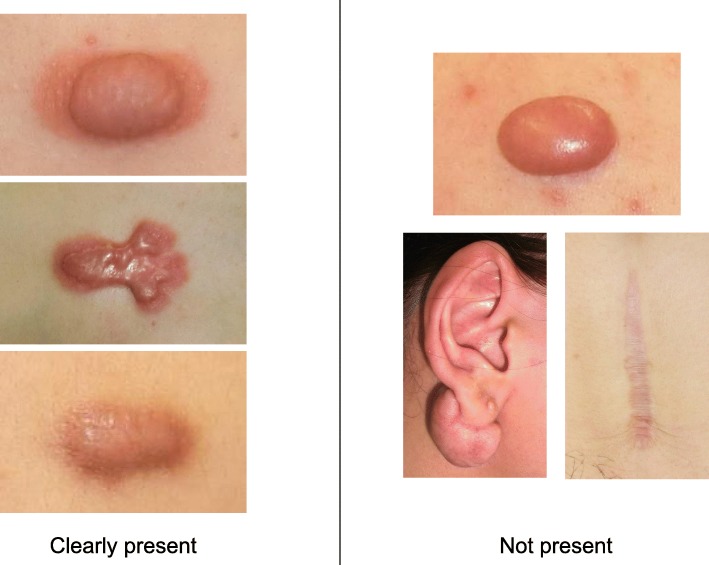
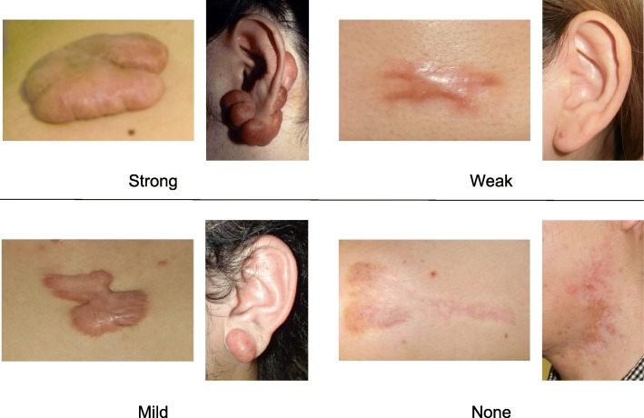
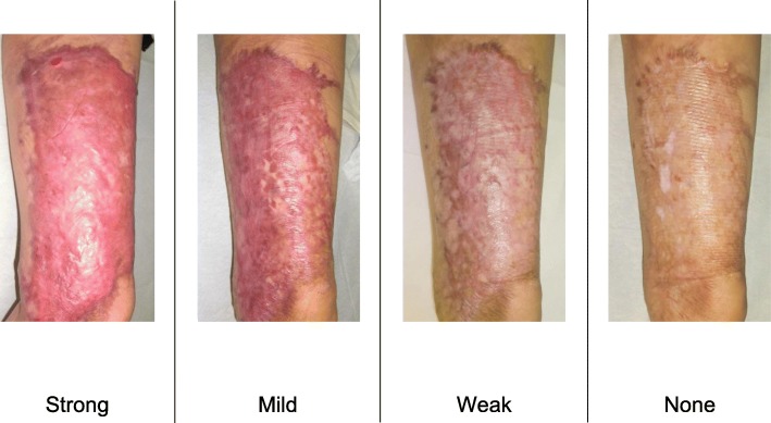
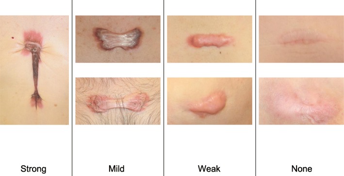
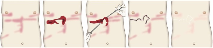
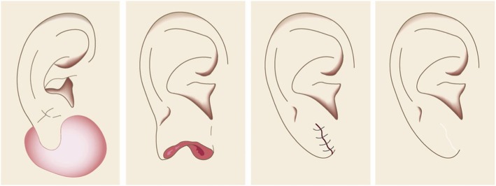
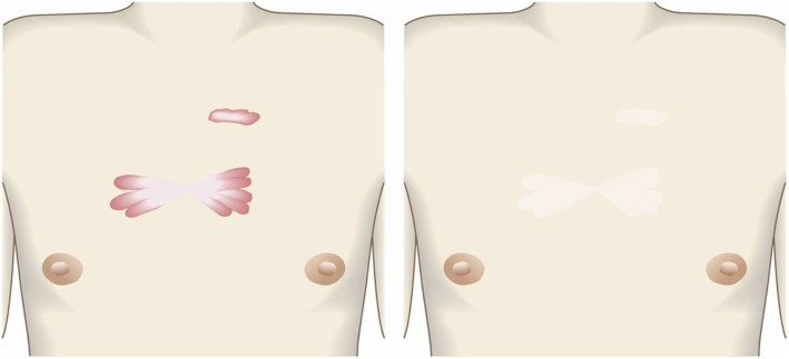
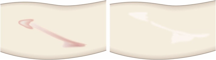
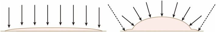
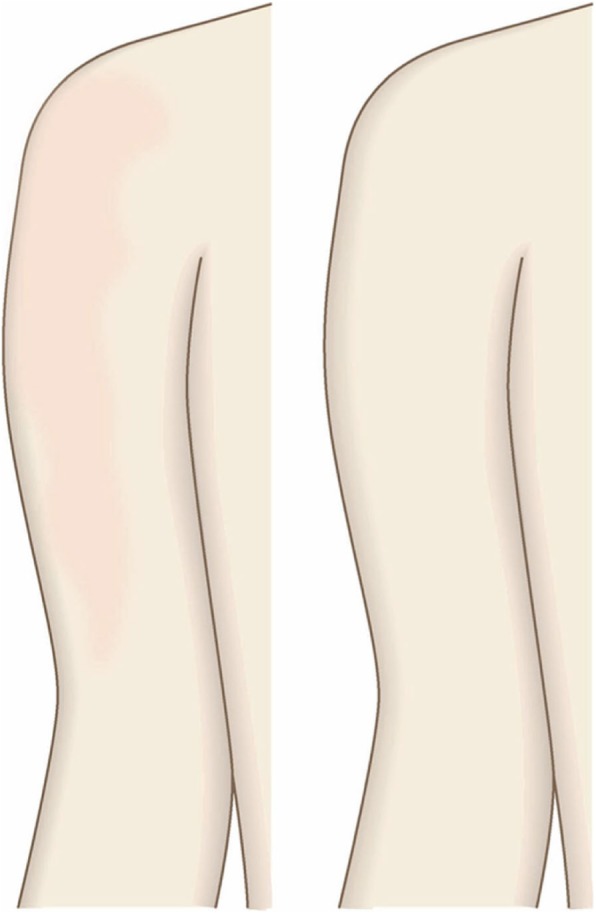
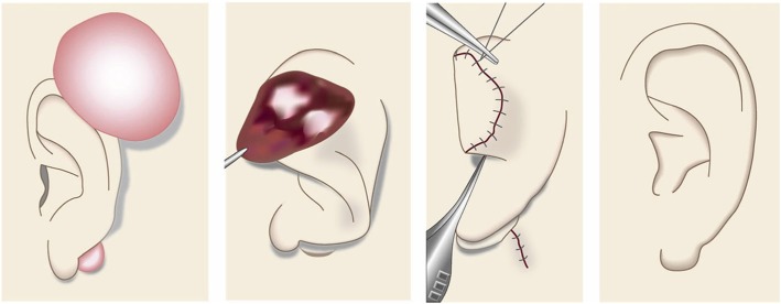
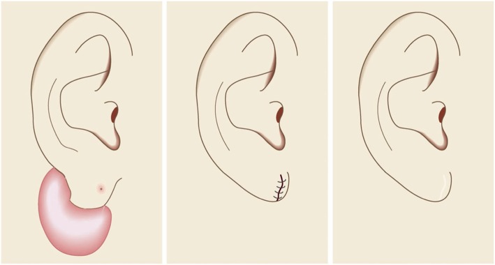
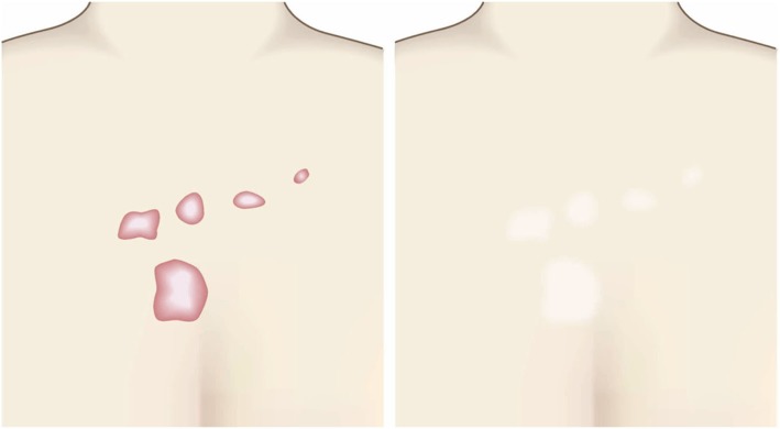
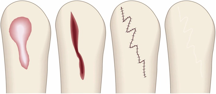
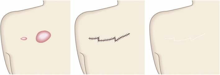
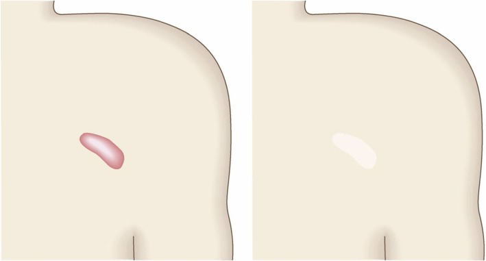
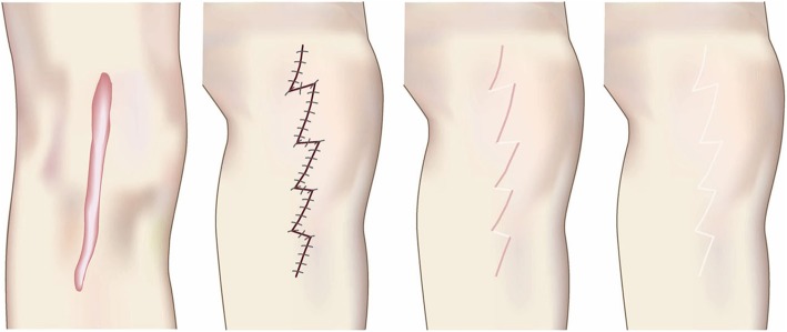
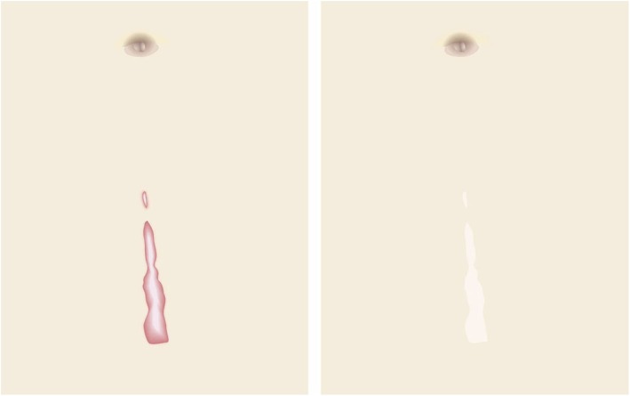
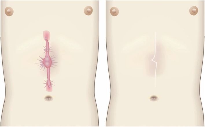
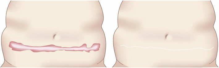
There has been a long-standing need for guidelines on the diagnosis and treatment of keloids and hypertrophic scars that are based on an understanding of the pathomechanisms that underlie these skin fibrotic diseases. This is particularly true for clinicians who deal with Asian and African patients because these ethnicities are highly prone to these diseases. By contrast, Caucasians are less likely to develop keloids and hypertrophic scars, and if they do, the scars tend not to be severe. This ethnic disparity also means that countries vary in terms of their differential diagnostic algorithms. The lack of clear treatment guidelines also means that primary care physicians are currently applying a hotchpotch of treatments, with uneven outcomes. To overcome these issues, the Japan Scar Workshop (JSW) has created a tool that allows clinicians to objectively diagnose and distinguish between keloids, hypertrophic scars, and mature scars. This tool is called the JSW Scar Scale (JSS) and it involves scoring the risk factors of the individual patients and the affected areas. The tool is simple and easy to use. As a result, even physicians who are not accustomed to keloids and hypertrophic scars can easily diagnose them and judge their severity. The JSW has also established a committee that, in cooperation with outside experts in various fields, has prepared a Consensus Document on keloid and hypertrophic scar treatment guidelines. These guidelines are simple and will allow even inexperienced clinicians to choose the most appropriate treatment strategy. The Consensus Document is provided in this article. It describes (1) the diagnostic algorithm for pathological scars and how to differentiate them from clinically similar benign and malignant tumors, (2) the general treatment algorithms for keloids and hypertrophic scars at different medical facilities, (3) the rationale behind each treatment for keloids and hypertrophic scars, and (4) the body site-specific treatment protocols for these scars. We believe that this Consensus Document will be helpful for physicians from all over the world who treat keloids and hypertrophic scars.

Keywords: Guideline; Hypertrophic scars; Keloid; Laser; Pathological scars; Pathology; Radiotherapy; Steroid; Surgery.


Conflict of interest statement

Competing interestsThe authors declare that they have no competing interests.

Figures

Fig. 1
Fig. 1
JSW Scar Scale (JSS)
Fig. 2
Fig. 2
Vertical growth (elevation)
Fig. 3
Fig. 3
Horizontal growth
Fig. 4
Fig. 4
Shape
Fig. 5
Fig. 5
Erythema around scars
Fig. 6
Fig. 6
Elevation
Fig. 7
Fig. 7
Redness of scars
Fig. 8
Fig. 8
Erythema around scars
Fig. 9
Fig. 9
Pseudolymphoma
Fig. 10
Fig. 10
Mixed tumor of the skin
Fig. 11
Fig. 11
Xanthogranuloma
Fig. 12
Fig. 12
Dermatofibrosarcoma protuberans (DFSP)
Fig. 13
Fig. 13
Squamous cell carcinoma (SCC)
Fig. 14
Fig. 14
Typical hypertrophic scar
Fig. 15
Fig. 15
Intermediate lesion
Fig. 16
Fig. 16
Typical keloid
Fig. 17
Fig. 17
Typical hypertrophic scar (HE staining)
Fig. 18
Fig. 18
Typical keloid (HE staining)
Fig. 19
Fig. 19
Keloid and hypertrophic scar treatment algorithm for pediatric patients
Fig. 20
Fig. 20
Keloid and hypertrophic scar treatment algorithm for adult patients
Fig. 21
Fig. 21
Topical adrenocortical hormone agent (administered via tape/plaster). When the corticosteroid tape/plaster therapy starts to improve the height and stiffness of the keloid/hypertrophic scar, the tape/plaster area being used, the affixation duration, and the intervals between fresh applications should be reduced gradually
Fig. 22
Fig. 22
Local adrenocortical hormone agent (administered by injection). Corticosteroid injections rapidly improve the symptoms of keloids and hypertrophic scars but their drawback is injection-induced pain. Means to prevent this pain should be implemented
Fig. 23
Fig. 23
The target of the injection. When injecting pathological scars with corticosteroid, do not inject the solid central fibrotic mass of the lesion because the drug will not infiltrate the tissue adequately. Moreover, the rising pressure induced by the injection may cause pain. Instead, penetrate the scar from its border with the normal skin. The target is the deepest part of the scar and/or the periphery of the scar, where the inflammation is particularly pronounced
Fig. 24
Fig. 24
Topical agents (corticosteroid and non-steroidal antiinflammatory drug [NSAID] preparations, heparinoid ointment, and silicone gels and creams). Treatment with topical preparations such as corticosteroid, NSAID, heparinoid, and silicone ointments, gels, and creams reduce inflammation. The goal is to induce scar maturation. However, the shape of the scar will remain after maturation
Fig. 25
Fig. 25
Oral medicines (tranilast, Saireito). It is recommended to use oral medicines when the patient has huge and/or multiple keloids or hypertrophic scars, since these conditions suggest the presence of a systemic risk factor
Fig. 26
Fig. 26
Rest/fixation therapy (administered by applying fixation tape or gel sheets). Fixation tape and gel sheets can reduce the tension on the pathological scar, thereby promoting scar maturation
Fig. 27
Fig. 27
Compression therapy (administered by applying bandages, supporters, garments, etc.). Compression therapy acts by placing pressure on the blood vessels around and in the keloid/hypertrophic scar. This reduces the blood flow in the lesion, which in turn suppresses scar inflammation and promotes scar maturation
Fig. 28
Fig. 28
Surgical excision and closure with simple sutures. When excising keloids or hypertrophic scars from body sites that have strong skin tension (e.g., the trunk), the fatty tissues should be removed along with the scar. The fasciae should then be undermined. Thereafter, the fasciae should be sutured so that the upper layers of the skin approximate each other closely. This makes it easy to place dermal sutures with minimal tension
Fig. 29
Fig. 29
The ideal suture method. a The scar should be removed along with the fatty tissue under the scar. b Undermine below the deep fascia of the muscle and then suture first the deep fasciae and then the superficial fasciae. c This suturing strategy causes the upper skin layers to attach to each other naturally. Dermal sutures can then be started
Fig. 30
Fig. 30
Surgical excision using the core excision method or partial resection. If the lesion is large or if total removal might result in significant deformity, it is recommended to remove only the fibrous core of the keloid/hypertrophic scar
Fig. 31
Fig. 31
Surgical excision followed by z-plasty. If the incision line used to excise a keloid/hypertrophic scar follows the predominant direction of skin tension, z-plasty should be applied. This will disperse the tension on the wound/scar
Fig. 32
Fig. 32
Computer simulation of the effect on wound tension. Computer simulation of the effect on wound tension when the directions of the incision and the predominant skin tension do and do not coincide. a When the incision line follows the direction of skin tension, the tension on the entire length of the wound will be high during the wound healing process (red color in the upper panel). b If the incision lies perpendicular to the direction of skin tension, the force will be dispersed along the wound and less tension will be placed on the wound (green color in the lower panel)
Fig. 33
Fig. 33
Surgical excision followed by reconstruction with skin grafts or flaps. If primary closure after keloid/hypertrophic scar excision cannot be performed with low tension, it is best to consider reconstruction with skin grafts or flaps. The procedure should be followed with postoperative adjuvant therapies such as radiotherapy
Fig. 34
Fig. 34
Postoperative radiotherapy. Body sites differ in terms of the postoperative radiotherapy protocol that is needed to prevent recurrence after keloid/hypertrophic scar excision. For example, earlobe keloid surgery should be followed with 10 Gy/2 fractions/2 days radiotherapy
Fig. 35
Fig. 35
Radiation monotherapy. Radiation monotherapy may be suitable for the few cases in which surgery will be difficult to perform. The radiation monotherapy can improve the severe pain and itch of the keloid/hypertrophic scar
Fig. 36
Fig. 36
Laser therapy. Laser therapy can improve the color of keloids and hypertrophic scars. Flat scars are particularly indicated for laser therapy
Fig. 37
Fig. 37
The ideal irradiation method of lasers. When performing laser therapy, the laser beam (arrows) should be held perpendicularly to the scar surface. This 90° orientation should be maintained as the beam is passed over the curvature of the scar. When irradiating the boundary of the scar (dashed arrows), it is permissible to irradiate some of the adjacent normal skin
Fig. 38
Fig. 38
Make-up therapy. Medical make-up therapy can temporarily improve the appearance of keloids, hypertrophic scars, and mature scars. This can improve the mental health of the patient
Fig. 39
Fig. 39
The core excision method for the cartilaginous part of the auricle. When excising keloids or hypertrophic scars from the cartilaginous part of the auricle, it is important to maintain the shape of the auricle. The core excision method is particularly suitable for this purpose
Fig. 40
Fig. 40
The wedge excision method for the earlobe. Many keloids and hypertrophic scars of the earlobe originate from the piercing hole. Most primary cases can be treated by wedge excision and simple suture, which maintains the shape of the ear lobe
Fig. 41
Fig. 41
Simple closure method for the lower jaw. Multiple keloids and hypertrophic scars on the lower jaw can be converted into linear mature scars by surgery and postoperative therapies such as radiotherapy and/or corticosteroid tape/plaster and injection therapy
Fig. 42
Fig. 42
Simple closure method for the anterior chest wall (the scars developed from a midline chest incision). Surgery and postoperative radiotherapy is indicated for a broadening keloid/hypertrophic scar that is growing from a midline chest incision
Fig. 43
Fig. 43
Z-plasties for the anterior chest wall (the scars developed from non-midline incisions or acne/folliculitis). Most of the keloids and hypertrophic scars that spread laterally on the anterior chest are due to acne/folliculitis and minor surgery. It is recommended to use z-plasty after excising these scars because this will disperse the horizontal skin tension on the wound
Fig. 44
Fig. 44
Conservative therapies for the anterior chest wall. Conservative therapies such as laser therapy will help keloids and hypertrophic scars on the anterior chest to mature. The scar shape will remain but it will be inconspicuous after the scar becomes mature
Fig. 45
Fig. 45
Laser therapy for the anterior chest wall. Conservative therapies such as laser therapy are particularly indicated for small keloids and hypertrophic scars on the anterior chest
Fig. 46
Fig. 46
Z-plasties for the upper arm. After excising a keloid or hypertrophic scar on the upper arm, it is recommended add z-plasty to disperse the skin tension on the wound
Fig. 47
Fig. 47
Z-plasties for the scapular area. If the wound left after excising a scapular keloid/hypertrophic scar is long, it should be closed with z-plasty to disperse the skin tension on the wound
Fig. 48
Fig. 48
Conservative therapies for the scapular area. It is recommended to treat small keloids and hypertrophic scars on the scapula with corticosteroid tape/plaster or injection therapy
Fig. 49
Fig. 49
Z-plasties for joint areas (the hand, elbow, knee, and foot). If the pathological scar on a joint is thin and runs in the direction in which the joint is extended, it is recommended to completely excise the scar and perform z-plasties to disperse the tension on the wound
Fig. 50
Fig. 50
Conservative therapies for the abdomen. Keloids and hypertrophic scars that arise from a midline abdominal incision can be treated with conservative therapies. However, the shape of the scar will remain
Fig. 51
Fig. 51
Z-plasties for the abdomen (the scars developed from an abdominal midline incision). If surgery is selected to treat a midline abdominal scar, it will yield a thin linear scar that is barely visible after maturation. If the scar is long, z-plasties should be applied to disperse the skin tension on the wound
Fig. 52
Fig. 52
Simple suture method for the abdomen (the scars developed from non-midline incisions). Non-midline abdominal scars can often be removed completely and sutured primarily because the abdomen has a relatively large amount of skin
Fig. 53
Fig. 53
Conservative therapies for the abdomen (the scars developed from non-midline incisions). Conservative therapies such as laser therapy may also be suitable for non-midline abdominal scars
Fig. 54
Fig. 54
Conservative therapies for the suprapubic area. If a keloid or hypertrophic scar on the suprapubic region is infected, surgery is the first choice of therapy
Fig. 55
Fig. 55
Other body areas. Although rare, keloids and hypertrophic scars can occur on the face, external genitals, and foot soles. Depending on the affected body region and lesion severity, conservative therapies and/or surgery followed by the appropriate radiotherapy protocol are recommended

References

    1. Ogawa R, Akaishi S, Hyakusoku H. Differential and exclusive diagnosis of diseases that resemble keloids and hypertrophic scars. Ann Plast Surg. 2009;62:660–664. doi: 10.1097/SAP.0b013e31817e9d67. - DOI - PubMed
    1. Ogawa R, Akaishi S, Akita S, Okabe K, Shimizu T, Sunaga A, et al. Japan Scar Workshop (JSW) Scar Scale 2015. Available online at; http://www.scar-keloid.com/pdf/JSW_scar_scale_2015_EN.pdf. Accessed 1 Oct 2019.
    1. Mustoe TA, Cooter RD, Gold MH, Hobbs FD, Ramelet AA, Shakespeare PG, et al. International advisory panel on scar management. International clinical recommendations on scar management. Plast Reconstr Surg. 2002;110:560–571. doi: 10.1097/00006534-200208000-00031. - DOI - PubMed
    1. Huang C, Akaishi S, Hyakusoku H, Ogawa R. Are keloid and hypertrophic scar different forms of the same disorder? A fibroproliferative skin disorder hypothesis based on keloid findings. Int Wound J. 2014;11:517–522. doi: 10.1111/j.1742-481X.2012.01118.x. - DOI - PMC - PubMed
    1. Ogawa R. Keloid and hypertrophic scars are the result of chronic inflammation in the reticular dermis. Int J Mol Sci. 2017;18:E606. doi: 10.3390/ijms18030606. - DOI - PMC - PubMed