

Autocrine/paracrine actions of growth hormone in human melanoma cell lines
- PMID: 31890904
- PMCID: PMC6928330
- DOI: 10.1016/j.bbrep.2019.100716
Autocrine/paracrine actions of growth hormone in human melanoma cell lines
Abstract
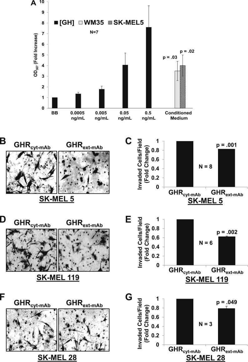
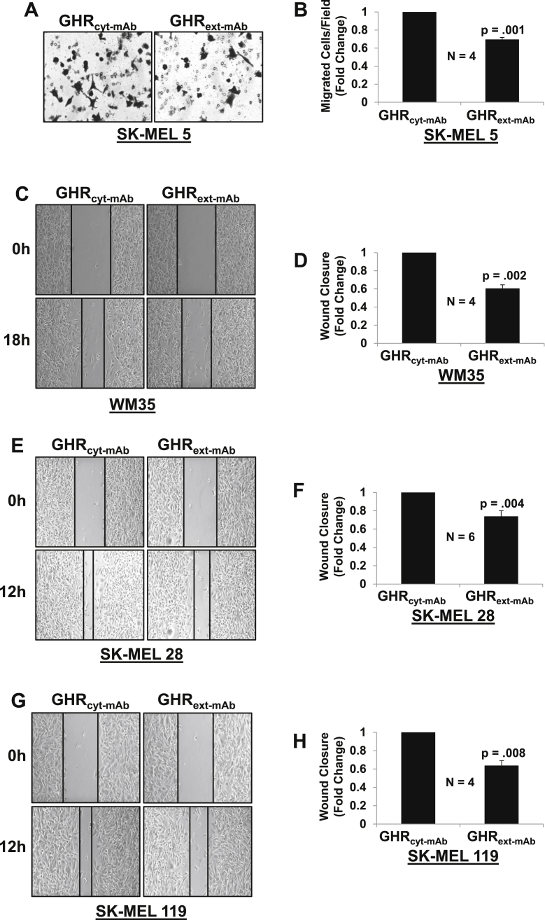
Melanoma is the most aggressive skin cancer. Its aggressiveness is most commonly attributed to ERK pathway mutations leading to constitutive signaling. Though initial tumor regression results from targeting this pathway, resistance often emerges. Interestingly, interrogation of the NCI-60 database indicates high growth hormone receptor (GHR) expression in melanoma cell lines. To further characterize melanoma, we tested responsiveness to human growth hormone (GH). GH treatment resulted in GHR signaling and increased invasion and migration, which was inhibited by a GHR monoclonal antibody (mAb) antagonist in WM35, SK-MEL 5, SK-MEL 28 and SK-MEL 119 cell lines. We also detected GH in the conditioned medium (CM) of human melanoma cell lines. GHR, JAK2 and STAT5 were basally phosphorylated in these cell lines, consistent with autocrine/paracrine GH production. Together, our results suggest that melanomas are enriched in GHR and produce GH that acts in an autocrine/paracrine manner. We suggest that GHR may constitute a therapeutic target in melanoma.
Keywords: Autocrine; Growth hormone (GH); Growth hormone receptor (GHR); Melanoma; Paracrine.
© 2019 The Authors.
Conflict of interest statement
The authors declare no potential conflicts of interest.
Figures


Similar articles
-
Human metastatic melanoma cell lines express high levels of growth hormone receptor and respond to GH treatment.Biochem Biophys Res Commun. 2013 Nov 8;441(1):144-50. doi: 10.1016/j.bbrc.2013.10.023. Epub 2013 Oct 14. Biochem Biophys Res Commun. 2013. PMID: 24134847 Free PMC article.
-
Caveolar and lipid raft localization of the growth hormone receptor and its signaling elements: impact on growth hormone signaling.J Biol Chem. 2004 May 14;279(20):20898-905. doi: 10.1074/jbc.M400625200. Epub 2004 Mar 8. J Biol Chem. 2004. PMID: 15010456
-
Growth hormone (GH) receptors in prostate cancer: gene expression in human tissues and cell lines and characterization, GH signaling and androgen receptor regulation in LNCaP cells.Mol Cell Endocrinol. 2004 May 31;220(1-2):109-23. doi: 10.1016/j.mce.2004.03.004. Mol Cell Endocrinol. 2004. PMID: 15196705
-
Morphogenic and tumorigenic potentials of the mammary growth hormone/growth hormone receptor system.Mol Cell Endocrinol. 2002 Nov 29;197(1-2):153-65. doi: 10.1016/s0303-7207(02)00259-9. Mol Cell Endocrinol. 2002. PMID: 12431808 Review.
-
Autocrine/paracrine roles of extrapituitary growth hormone and prolactin in health and disease: An overview.Gen Comp Endocrinol. 2015 Sep 1;220:103-11. doi: 10.1016/j.ygcen.2014.11.004. Epub 2014 Nov 15. Gen Comp Endocrinol. 2015. PMID: 25448258 Review.
Cited by
-
Melanoma-Keratinocyte Crosstalk Participates in Melanoma Progression with Mechanisms Partially Overlapping with Those of Cancer-Associated Fibroblasts.Int J Mol Sci. 2025 Aug 15;26(16):7901. doi: 10.3390/ijms26167901. Int J Mol Sci. 2025. PMID: 40869222 Free PMC article.
-
Approach of Multiple Endocrine Neoplasia Type 1 (MEN1) Syndrome-Related Skin Tumors.Diagnostics (Basel). 2022 Nov 12;12(11):2768. doi: 10.3390/diagnostics12112768. Diagnostics (Basel). 2022. PMID: 36428828 Free PMC article. Review.
-
Growth hormone receptor promotes breast cancer progression via the BRAF/MEK/ERK signaling pathway.FEBS Open Bio. 2020 Jun;10(6):1013-1020. doi: 10.1002/2211-5463.12816. Epub 2020 May 6. FEBS Open Bio. 2020. PMID: 32069380 Free PMC article.
-
Melanoma-derived cytokines and extracellular vesicles are interlinked with macrophage immunosuppression.Front Mol Biosci. 2025 Jan 22;11:1522717. doi: 10.3389/fmolb.2024.1522717. eCollection 2024. Front Mol Biosci. 2025. PMID: 39911494 Free PMC article.
-
Synergistic effects of immunoregulation and osteoinduction of ds-block elements on titanium surface.Bioact Mater. 2020 Aug 14;6(1):191-207. doi: 10.1016/j.bioactmat.2020.08.001. eCollection 2021 Jan. Bioact Mater. 2020. PMID: 32913928 Free PMC article.
References
-
- An S., Yang Y., Ward R., Liu Y., Guo X.X., Xu T.R., A-Raf A new star of the family of raf kinases. Crit. Rev. Biochem. Mol. Biol. 2015;50:520–531. - PubMed
-
- Chapman P.B. Mechanisms of resistance to RAF inhibition in melanomas harboring a BRAF mutation. Am. Soc. Clin. Oncol. Educ. Book. 2013 - PubMed
-
- Isaksson O.G., Eden S., Jansson J.O. Mode of action of pituitary growth hormone on target cells. Annu. Rev. Physiol. 1985;47:483–499. - PubMed
Grants and funding
LinkOut - more resources
Full Text Sources
Miscellaneous
