. 2019 Dec 17;5(12):e02943.
doi: 10.1016/j.heliyon.2019.e02943. eCollection 2019 Dec.

Substrate specificity of thioredoxins and glutaredoxins - towards a functional classification

Affiliations

Substrate specificity of thioredoxins and glutaredoxins - towards a functional classification

Manuela Gellert et al. Heliyon. .

Abstract

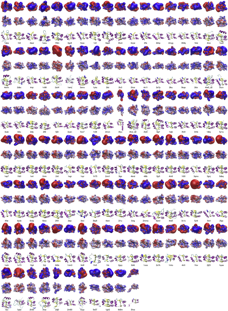
The spatio-temporal reduction and oxidation of protein thiols is an essential mechanism in signal transduction in all kingdoms of life. Thioredoxin (Trx) family proteins efficiently catalyze thiol-disulfide exchange reactions and the proteins are widely recognized for their importance in the operation of thiol switches. Trx family proteins have a broad and at the same time very distinct substrate specificity - a prerequisite for redox switching. Despite of multiple efforts, the true nature for this specificity is still under debate. Here, we comprehensively compare the classification/clustering of various redoxins from all domains of life based on their similarity in amino acid sequence, tertiary structure, and their electrostatic properties. We correlate these similarities to the existence of common interaction partners, identified in various previous studies and suggested by proteomic screenings. These analyses confirm that primary and tertiary structure similarity, and thereby all common classification systems, do not correlate to the target specificity of the proteins as thiol-disulfide oxidoreductases. Instead, a number of examples clearly demonstrate the importance of electrostatic similarity for their target specificity, independent of their belonging to the Trx or glutaredoxin subfamilies.

Keywords: Biocomputational method; Biomolecules; Electrostatics; Glutaredoxin; Gromov-Wasserstein distance; Mathematical biosciences; Molecular docking; Protein-protein interaction; Redox signaling; Thioredoxin.


Figures

Figure 1
Figure 1
Clustering of human redoxins. (A) Phylogram based on primary structure comparison, computed by Clustal Omega and CLC sequence viewer. (B) Similarity tree based on the similarity of the 3D structures extracted from the pdb and generated by homology modeling; the tree was computed using UCSF Chimera and the CLC sequence viewer. (C) The electrostatic similarity of the whole proteins was computed as outlined in the methods section; the tree was generated using ‘R’. The protein abbreviations highlighted in green are referred to in the main text. The Trx proteins with a Cys-Gly-Pro-Cys active site motif were highlighted with a red circle in B.
Figure 2
Figure 2
Electrostatic features of the active site contact areas of the human redoxins. The first rows depict the electrostatic potential isosurfaces at +/- 1 K T·e−1. The second row depicts the electrostatic potential at +/- 4 K T·e−1 mapped to the water-accessible surface of the proteins. Blue: positive, red negative potential. The third row depicts the proteins in cartoon models, helices are colored in purple, sheets in yellow. The proteins were arranged with the N-terminal active site thiol in the middle of the models. The electrostatic similarity of the whole proteins was computed as outlined in the methods section.
Figure 3
Figure 3
Common interaction partners between the human redoxins. (A) Pair-wise comparison between all human redoxins. The total numbers of potential interactions partners collected from various data sources is depicted with gray background in the diagonal; yellow background: 3–4 common interaction partners; light green background: 5–9 common interaction partners; green background: ≥ 10 common interaction partners. The full list of interaction partners can be found in the supplementary tables. (B–C) Venn diagrams of the overlapping potential interactions partners between Trx1, Nrx, Txndc9, Txndc17, and Txnl1 (B) as well as Grx1, Grx2, Grx3, and Grx5 (B).
Figure 4
Figure 4
Clustering of all representative redoxins in the pdb. (A) Phylogram based on primary structure comparison, computed by Clustal Omega and CLC sequence viewer. The dashed red line separates the Trx and Grx subfamilies. (B) The electrostatic similarity of the whole proteins was computed as outlined in the methods section; the tree was generated using ‘R’. The red asterisks mark proteins interacting with E. coli RNR, the black asterisks proteins interacting with E. coli PAPS reductase. The color code is included in the figure. Further information on the protein structures can be obtained from the supplementary Table, sheet 5.
Figure 5
Figure 5
Electrostatic features of all representative redoxins in the pdb. The first rows depict the electrostatic potential isosurfaces at +/- 1 K T·e−1. The second row depicts the electrostatic potential at +/- 4 K T·e−1 mapped to the water-accessible surface of the proteins. Blue: positive, red negative potential. The third row depicts the proteins in cartoon models, helices are colored in purple, sheets in yellow. The pdb entry code of the structures is indicated in the fourth row. The proteins were arranged with the N-terminal active site thiol in the middle of the models. The electrostatic similarity of the whole proteins was computed as outlined in the methods section.

Similar articles

Cited by

References

    1. Ghezzi P., Bonetto V., Fratelli M. Thiol disulfide balance: from the concept of oxidative stress to that of redox regulation. Antioxidants Redox Signal. 2005;7:964–972. - PubMed
    1. Janssen-Heininger Y.M.W., Mossman B.T., Heintz N.H., Forman H.J., Kalyanaraman B., Finkel T., Stamler J.S., Rhee S.G., van der Vliet A. Redox-based regulation of signal transduction: principles, pitfalls, and promises. Free Radic. Biol. Med. 2008;45:1–17. - PMC - PubMed
    1. Drazic A., Winter J. The physiological role of reversible methionine oxidation. Biochim. Biophys. Acta. 2014;1844:1367–1382. - PubMed
    1. Hanschmann E.-M., Godoy J.R., Berndt C., Hudemann C., Lillig C.H. Thioredoxins, glutaredoxins, and peroxiredoxins--molecular mechanisms and health significance: from cofactors to antioxidants to redox signaling. Antioxidants Redox Signal. 2013;19:1539–1605. - PMC - PubMed
    1. Martin J.L. Thioredoxin--a fold for all reasons. Struct. Lond. Engl. 1993. 1995;3:245–250. - PubMed

LinkOut - more resources