. 2019 Dec 10;4(26):21891-21899.
doi: 10.1021/acsomega.9b02883. eCollection 2019 Dec 24.

Serum Albumin-Peptide Conjugates for Simultaneous Heparin Binding and Detection

Affiliations

Serum Albumin-Peptide Conjugates for Simultaneous Heparin Binding and Detection

Qing Liu et al. ACS Omega. .

Abstract

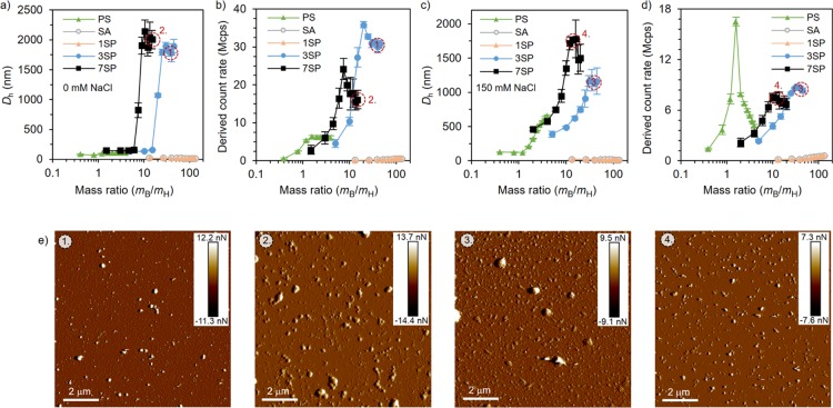
Heparin is a polysaccharide-based anticoagulant agent, which is widely used in surgery and blood transfusion. However, overdosage of heparin may cause severe side effects such as bleeding and low blood platelet count. Currently, there is only one clinically licensed antidote for heparin: protamine sulfate, which is known to provoke adverse effects. In this work, we present a stable and biocompatible alternative for protamine sulfate that is based on serum albumin, which is conjugated with a variable number of heparin-binding peptides. The heparin-binding efficiency of the conjugates was evaluated with methylene blue displacement assay, dynamic light scattering, and anti-Xa assay. We found that multivalency of the peptides played a key role in the observed heparin-binding affinity and complex formation. The conjugates had low cytotoxicity and low hemolytic activity, indicating excellent biocompatibility. Furthermore, a sensitive DNA competition assay for heparin detection was developed. The detection limit of heparin was 0.1 IU/mL, which is well below its therapeutic range (0.2-0.4 IU/mL). Such biomolecule-based systems are urgently needed for next-generation biocompatible materials capable of simultaneous heparin binding and sensing.


Conflict of interest statement

The authors declare no competing financial interest.

Figures

Figure 1
Figure 1
(a) Schematic presentation of the preparation of single (1SP) and multiple (3SP and 7SP) peptide-modified SAs. (b) MALDI-TOF spectra of SA and 1SP: MSA = 66,519 g/mol and M1SP = 68,414 g/mol (theoretical molecular weights: MSA = 66,430 g/mol and M1SP = 68,636 g/mol); (c) MALDI-TOF spectra of cross-linker-capped SAs: M = 67,502 g/mol and M10× = 68,473 g/mol. Therefore, the estimated number of cross-linkers attached to SAs for 5 and 10 equiv syntheses was 3 and 7, respectively; (d) HPLC analysis of purified 1SP, 3SP, and 7SP using a heparin affinity column. SA was analyzed for comparison.
Figure 2
Figure 2
MB displacement assay results in (left) mass and (right) molar ratios of the binder to heparin, measured by the A (664 nm)/A (568 nm) ratio. Heparin concentration: 0.017 mg/mL (0.9 μM). Protamine sulfate was plotted for comparison. Measurements were performed using triplicate samples, and the averaged results with standard deviation are presented.
Figure 3
Figure 3
Titration of heparin (0.02 mg/mL) with binders (a, b) in the 2 mM Tris-HCl buffer and (c, d) at the physiological salt concentration (2 mM Tris-HCl, 150 mM NaCl) was monitored with DLS. The hydrodynamic diameter (a, c) and derived count rate (b, d) were used as indicators for the successful complexation. Titrations were performed using triplicate samples, and the averaged results with standard deviation are presented. (e) Peak force tapping mode AFM images (insets show the peak force error scale) of heparin neutralized with (1, 3) 3SP (mB/mH = 40) and (2, 4) 7SP (mB/mH = 15) in the presence of 0 mM (1, 2) and 150 mM (3, 4) NaCl. The corresponding DLS samples have been marked on the data.
Figure 4
Figure 4
Heparin neutralization measured with the anti-Xa assay in plasma. Experiments were performed using triplicate samples, and the averaged results with standard deviation are presented.
Figure 5
Figure 5
Effect of the heparin binders on cell viability evaluated with MTT assay using HDF cells. (left) Cells were incubated with free binders at four concentrations for 24 h. (right) Heparin of 1 μg/mL was first neutralized by binders and then incubated with the cells for 4, 14, or 24 h. Measurements were performed using triplicate samples, and the averaged results with standard deviation are presented.
Figure 6
Figure 6
Hemolysis assay with RBCs; results indicate that both (a) binders alone and (b) binder–heparin (5 μg/mL) complexes cause negligible red blood cell hemolysis. Solutions of 1× phosphate-buffered saline (PBS) and 1% Triton X-100 were used as the negative and positive control, respectively. Measurements were performed using triplicate samples, and the averaged results with standard deviation are presented.
Figure 7
Figure 7
Fluorescence switch-on detection of heparin in the 2 mM Tris-HCl buffer with the 7SP–DNA complex. Blue squares (on the left-hand side) show the titration of 0.2 μM DNA with 7SP. Orange triangles (on the right-hand side) show the fluorescence recovered upon adding heparin to the solution of the 7SP–DNA complex. The insets show schematic steps of the process. Titrations were performed using triplicate samples, and the averaged results with standard deviation are presented.

References

    1. Casu B.; Naggi A.; Torri G. Re-Visiting the Structure of Heparin. Carbohydr. Res. 2015, 403, 60–68. 10.1016/j.carres.2014.06.023. - DOI - PubMed
    1. Bromfield S. M.; Wilde E.; Smith D. K. Heparin Sensing and Binding-Taking Supramolecular Chemistry Towards Clinical Applications. Chem. Soc. Rev. 2013, 42, 9184–9195. 10.1039/c3cs60278h. - DOI - PubMed
    1. Esko J. D.; Selleck S. B. Order Out of Chaos: Assembly of Ligand Binding Sites in Heparin Sulfate. Annu. Rev. Biochem. 2002, 71, 435–471. 10.1146/annurev.biochem.71.110601.135458. - DOI - PubMed
    1. Hirsh J.; Raschke R. Heparin and Low-Molecular-Weight Heparin: The Seventh ACCP Conference on Antithrombotic and Thrombolytic Therapy. Chest 2004, 126, 188S–203S. 10.1378/chest.126.3_suppl.188S. - DOI - PubMed
    1. Warkentin T. E.; Levine M. N.; Hirsh J.; Horsewood P.; Roberts R. S.; Gent M.; Kelton J. G. Heparin-Induced Thrombocytopenia in Patients Treated with Low-Molecular-Weight Heparin or Unfractionated Heparin. N. Engl. J. Med. 1995, 332, 1330–1336. 10.1056/NEJM199505183322003. - DOI - PubMed

LinkOut - more resources