

Spatial distribution of type II collagen gene expression in the mouse intervertebral disc
- PMID: 31891119
- PMCID: PMC6920692
- DOI: 10.1002/jsp2.1070
Spatial distribution of type II collagen gene expression in the mouse intervertebral disc
Abstract
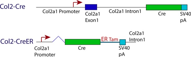
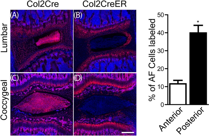
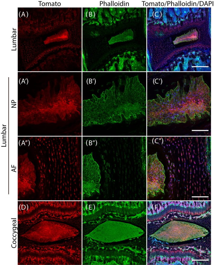
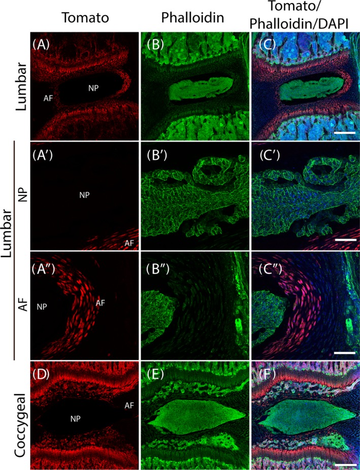
Genetic tools such as the Cre-Lox reporter system are powerful aids for tissue-specific cell tracking. For example, it would be useful in examining intervertebral disc (IVD) cell populations in normal and diseased states. A Cre recombinase and its recognition site, loxP have been adapted from the bacteriophage for use in genetic manipulation. The reporter mice used here express the red fluorescent protein, tdTomato with flanking LoxP sites (Rosa26 TdTomato mice). We compared two different Collagen type II (Col2) promoter constructs that drive Cre-recombinase expression in mice: (a) Col2-Cre, which allows constitutive Cre-recombinase expression under the control of the Col2 promoter/enhancer and (b) Col2-CreER, which contains a shorter promoter/enhancer region than Col2-Cre, but has human estrogen binding elements that bind tamoxifen, resulting in Cre-recombinase expression. The goal of the study is to characterize Cre-recombinase distribution pattern in Col2-Cre and Col2-CreER mice using tdTomato as reporter in the spine. The expression patterns of these two mice were further compared with Col2 gene expression in the native mouse NP and AF tissues by real-time PCR. We crossed Col2-Cre mice or Col2-CreER mice with the tdTomato reporter mice, and compared the tdTomato expression patterns. Col2-CreER/tdTomato mice were injected with tamoxifen at postnatal day 7 to activate the Cre-recombinase. TdTomato in the constitutively active Col2-Cre mice was detected in the nucleus pulposus (NP), the entire annulus fibrosus (AF), and in cartilaginous endplate and growth plate cells in the lower lumbar and coccygeal spine. In contrast, when Col2-CreER activity was induced by tamoxifen at P7, tdTomato was limited to the inner AF, and was absent from the NP. We have described the differences in Col2 reporter gene expression, in Col2-Cre/tdTomato and Col2-Cre-ER/tdTomato mouse IVD. The information provided here will help to guide future investigations of IVD biology.
Keywords: Cre‐Lox reporter system; gene regulation; intervertebral disc; mouse model; type II collagen (Col2).
© 2019 The Authors. JOR Spine published by Wiley Periodicals, Inc. on behalf of Orthopaedic Research Society.
Conflict of interest statement
The authors declare no competing conflict of interest.
Figures
Similar articles
-
Type II collagen-positive embryonic progenitors are the major contributors to spine and intervertebral disc development and repair.Stem Cells Transl Med. 2021 Oct;10(10):1419-1432. doi: 10.1002/sctm.20-0424. Epub 2021 May 25. Stem Cells Transl Med. 2021. PMID: 34032373 Free PMC article.
-
Characterization of Cre recombinase mouse lines enabling cell type-specific targeting of postnatal intervertebral discs.J Cell Physiol. 2019 Sep;234(9):14422-14431. doi: 10.1002/jcp.28166. Epub 2019 Jan 23. J Cell Physiol. 2019. PMID: 30675722 Free PMC article.
-
Col2-Cre and tamoxifen-inducible Col2-CreER target different cell populations in the knee joint.Osteoarthritis Cartilage. 2016 Jan;24(1):188-91. doi: 10.1016/j.joca.2015.07.025. Epub 2015 Aug 6. Osteoarthritis Cartilage. 2016. PMID: 26256767 Free PMC article.
-
Establishment of Neh2-Cre:tdTomato reporter mouse for monitoring the exposure history to electrophilic stress.Free Radic Biol Med. 2022 Nov 20;193(Pt 2):610-619. doi: 10.1016/j.freeradbiomed.2022.11.004. Epub 2022 Nov 9. Free Radic Biol Med. 2022. PMID: 36368569 Review.
-
Conditional gene expression in the mouse inner ear using Cre-loxP.J Assoc Res Otolaryngol. 2012 Jun;13(3):295-322. doi: 10.1007/s10162-012-0324-5. Epub 2012 Apr 24. J Assoc Res Otolaryngol. 2012. PMID: 22526732 Free PMC article. Review.
Cited by
-
Type II collagen-positive embryonic progenitors are the major contributors to spine and intervertebral disc development and repair.Stem Cells Transl Med. 2021 Oct;10(10):1419-1432. doi: 10.1002/sctm.20-0424. Epub 2021 May 25. Stem Cells Transl Med. 2021. PMID: 34032373 Free PMC article.
-
Intervertebral Disc-on-a-ChipMF: A New Model for Mouse Disc Culture via Integrating Mechanical Loading and Dynamic Media Flow.Adv Mater Technol. 2023 Nov 10;8(21):2300606. doi: 10.1002/admt.202300606. Epub 2023 Aug 27. Adv Mater Technol. 2023. PMID: 39130370 Free PMC article.
-
Constitutive and conditional gene knockout mice for the study of intervertebral disc degeneration: Current status, decision considerations, and future possibilities.JOR Spine. 2023 Jan 7;6(1):e1242. doi: 10.1002/jsp2.1242. eCollection 2023 Mar. JOR Spine. 2023. PMID: 36994464 Free PMC article. Review.
-
Elevated inflammatory gene expression in intervertebral disc tissues in mice with ADAM8 inactivated.Sci Rep. 2021 Jan 19;11(1):1804. doi: 10.1038/s41598-021-81495-y. Sci Rep. 2021. PMID: 33469101 Free PMC article.
-
N6-Methyladenosine-Modified circSMAD4 Prevents Lumbar Instability Induced Cartilage Endplate Ossification.Adv Sci (Weinh). 2025 Apr;12(13):e2413970. doi: 10.1002/advs.202413970. Epub 2025 Feb 12. Adv Sci (Weinh). 2025. PMID: 39936497 Free PMC article.
References
-
- Eyre DR, Muir H. Quantitative analysis of types I and II collagens in human intervertebral discs at various ages. Biochim Biophys Acta. 1977;492:29‐42. - PubMed
Grants and funding
LinkOut - more resources
Full Text Sources
Molecular Biology Databases
Miscellaneous
