

Detection of Bartonella spp. in dogs after infection with Rickettsia rickettsii
- PMID: 31891215
- PMCID: PMC6979086
- DOI: 10.1111/jvim.15675
Detection of Bartonella spp. in dogs after infection with Rickettsia rickettsii
Abstract
Background: Dynamics of infection by Bartonella and Rickettsia species, which are epidemiologically associated in dogs, have not been explored in a controlled setting.
Objectives: Describe an outbreak investigation of occult Bartonella spp. infection among a group of dogs, discovered after experimentally induced Rickettsia rickettsii (Rr) infection.
Animals: Six apparently healthy purpose-bred Beagles obtained from a commercial vendor.
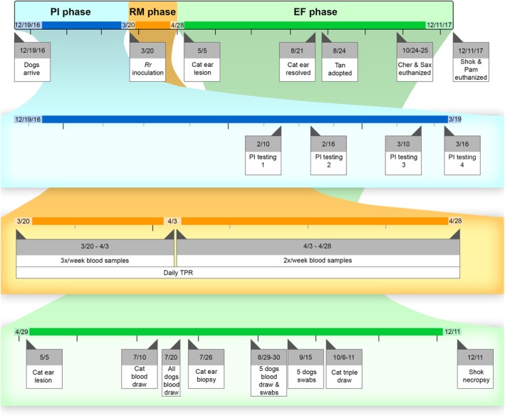
Methods: Retrospective and prospective study. Dogs were serially tested for Bartonella spp. and Rr using serology, culture, and PCR, over 3 study phases: 3 months before inoculation with Rr (retrospective), 6 weeks after inoculation with Rr (retrospective), and 8 months of follow-up (prospective).
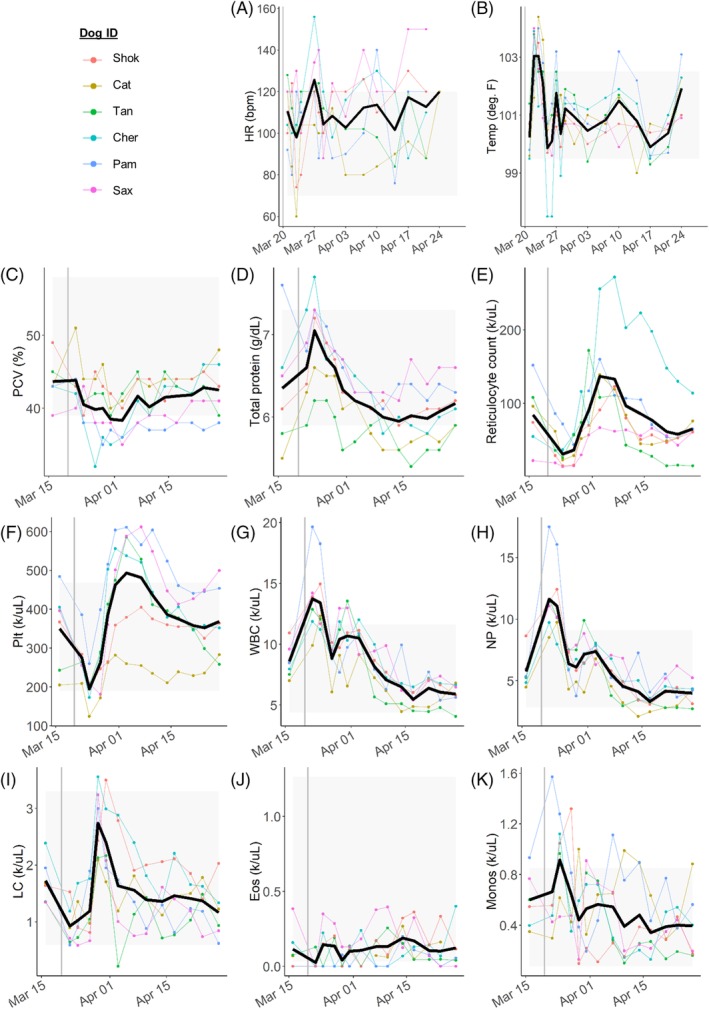
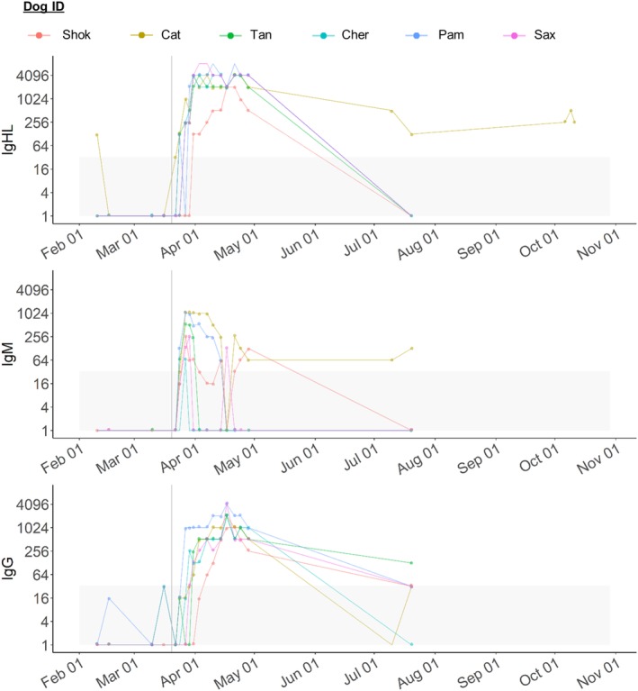
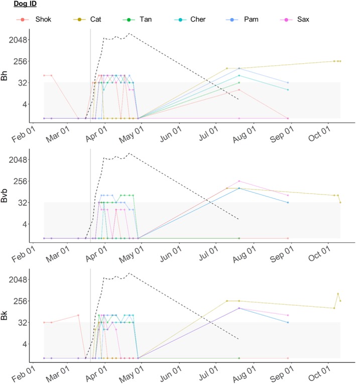
Results: Before Rr infection, 1 dog was Bartonella henselae (Bh) immunofluorescent antibody assay (IFA) seroreactive and 1 was Rickettsia spp. IFA seroreactive. After inoculation with Rr, all dogs developed mild Rocky Mountain spotted fever compatible with low-dose Rr infection, seroconverted to Rickettsia spp. within 4-11 days, and recovered within 1 week. When 1 dog developed ear tip vasculitis with intra-lesional Bh, an investigation of Bartonella spp. infection was undertaken. All dogs had seroconverted to 1-3 Bartonella spp. between 7 and 18 days after Rr inoculation. Between 4 and 8 months after Rr inoculation, Bh DNA was amplified from multiple tissues from 2 dogs, and Bartonella vinsonii subsp. berkhoffii (Bvb) DNA was amplified from 4 of 5 dogs' oral swabs.
Conclusions and clinical importance: Vector-borne disease exposure was demonstrated in research dogs from a commercial vendor. Despite limitations, our results support the possibilities of recrudescence of chronic subclinical Bartonella spp. infection after Rr infection and horizontal direct-contact transmission between dogs.
Keywords: PCR; recrudescence; serology; transmission.
© 2019 The Authors. Journal of Veterinary Internal Medicine published by Wiley Periodicals, Inc. on behalf of the American College of Veterinary Internal Medicine.
Conflict of interest statement
In conjunction with Dr. S. Sontakke and North Carolina State University, E. B. Breitschwerdt holds US Patent No. 7,115,385; Media and Methods for Cultivation of Microorganisms, which was issued on October 3, 2006. He is a co‐founder, shareholder and Chief Scientific Officer for Galaxy Diagnostics, a company that provides advanced diagnostic testing for the detection of Bartonella spp. infections. Ramaswamy Chandrashekar is employed by IDEXX Laboratories. The remaining authors declare no conflicts of interest.
Figures
Similar articles
-
Evaluation of cell culture-grown Bartonella antigens in immunofluorescent antibody assays for the serological diagnosis of bartonellosis in dogs.J Vet Intern Med. 2018 Nov;32(6):1958-1964. doi: 10.1111/jvim.15301. Epub 2018 Oct 11. J Vet Intern Med. 2018. PMID: 30307643 Free PMC article.
-
Analysis of seroreactivity against cell culture-derived Bartonella spp. antigens in dogs.J Vet Intern Med. 2014 Jan-Feb;28(1):38-41. doi: 10.1111/jvim.12263. Epub 2013 Dec 16. J Vet Intern Med. 2014. PMID: 24341682 Free PMC article.
-
Experimental infection of dogs with Bartonella henselae and Bartonella vinsonii subsp. berkhoffii.Vet Immunol Immunopathol. 2013 Nov 15;156(1-2):153-8. doi: 10.1016/j.vetimm.2013.09.007. Epub 2013 Sep 21. Vet Immunol Immunopathol. 2013. PMID: 24120155
-
Neurobartonelloses: emerging from obscurity!Parasit Vectors. 2024 Oct 5;17(1):416. doi: 10.1186/s13071-024-06491-3. Parasit Vectors. 2024. PMID: 39369199 Free PMC article. Review.
-
An Update on the Laboratory Diagnosis of Rickettsia spp. Infection.Pathogens. 2021 Oct 13;10(10):1319. doi: 10.3390/pathogens10101319. Pathogens. 2021. PMID: 34684267 Free PMC article. Review.
Cited by
-
Bartonella spp. Prevalence (Serology, Culture, and PCR) in Sanitary Workers in La Rioja Spain.Pathogens. 2020 Mar 4;9(3):189. doi: 10.3390/pathogens9030189. Pathogens. 2020. PMID: 32143533 Free PMC article.
-
Experimental Infection of Ferrets with Bartonella henselae: In Search of a Novel Animal Model for Zoonotic Bartonellosis.Pathogens. 2025 Apr 26;14(5):421. doi: 10.3390/pathogens14050421. Pathogens. 2025. PMID: 40430742 Free PMC article.
-
Antibiotic Susceptibility of Bartonella Grown in Different Culture Conditions.Pathogens. 2021 Jun 8;10(6):718. doi: 10.3390/pathogens10060718. Pathogens. 2021. PMID: 34201011 Free PMC article.
-
Exposure of Domestic Cats to Three Zoonotic Bartonella Species in the United States.Pathogens. 2021 Mar 17;10(3):354. doi: 10.3390/pathogens10030354. Pathogens. 2021. PMID: 33802644 Free PMC article.
-
Bartonella spp. seroepidemiology and associations with clinicopathologic findings in dogs in the United States.J Vet Intern Med. 2022 Jan;36(1):116-125. doi: 10.1111/jvim.16311. Epub 2021 Nov 17. J Vet Intern Med. 2022. PMID: 34788481 Free PMC article.
References
-
- Breitschwerdt EB. Bartonellosis, one health and all creatures great and small. Vet Dermatol. 2017;28(1):96‐e21. - PubMed
-
- Breitschwerdt E. Bartonellosis: one health perspectives for an emerging infectious disease. ILAR J. 2014;55(1):46‐58. - PubMed
-
- Golly E, Breitschwerdt EB, Balakrishnan N, et al. Bartonella henselae, Bartonella koehlerae, and Rickettsia rickettsii seroconversion and seroreversion in a dog with acute‐onset fever, lameness, and lymphadenopathy followed by a protracted disease course. Vet Parasitol Reg Stud Report. 2016;7:19‐24. - PubMed
-
- Breitschwerdt EB, Blann KR, Stebbins ME, et al. Clinicopathological abnormalities and treatment response in 24 dogs seroreactive to Bartonella vinsonii (berkhoffii) antigens. J Am Anim Hosp Assoc. 2004;40(2):92‐101. - PubMed
MeSH terms
Grants and funding
LinkOut - more resources
Full Text Sources
Miscellaneous
