. 2020 May;9(5):620-635.
doi: 10.1002/sctm.19-0277. Epub 2019 Dec 31.

Adaptation within embryonic and neonatal heart environment reveals alternative fates for adult c-kit+ cardiac interstitial cells

Affiliations

Adaptation within embryonic and neonatal heart environment reveals alternative fates for adult c-kit+ cardiac interstitial cells

Bingyan J Wang et al. Stem Cells Transl Med. 2020 May.

Abstract

Cardiac interstitial cells (CICs) perform essential roles in myocardial biology through preservation of homeostasis as well as response to injury or stress. Studies of murine CIC biology reveal remarkable plasticity in terms of transcriptional reprogramming and ploidy state with important implications for function. Despite over a decade of characterization and in vivo utilization of adult c-Kit+ CIC (cCIC), adaptability and functional responses upon delivery to adult mammalian hearts remain poorly understood. Limitations of characterizing cCIC biology following in vitro expansion and adoptive transfer into the adult heart were circumvented by delivery of the donated cells into early cardiogenic environments of embryonic, fetal, and early postnatal developing hearts. These three developmental stages were permissive for retention and persistence, enabling phenotypic evaluation of in vitro expanded cCICs after delivery as well as tissue response following introduction to the host environment. Embryonic blastocyst environment prompted cCIC integration into trophectoderm as well as persistence in amniochorionic membrane. Delivery to fetal myocardium yielded cCIC perivascular localization with fibroblast-like phenotype, similar to cCICs introduced to postnatal P3 heart with persistent cell cycle activity for up to 4 weeks. Fibroblast-like phenotype of exogenously transferred cCICs in fetal and postnatal cardiogenic environments is consistent with inability to contribute directly toward cardiogenesis and lack of functional integration with host myocardium. In contrast, cCICs incorporation into extra-embryonic membranes is consistent with fate of polyploid cells in blastocysts. These findings provide insight into cCIC biology, their inherent predisposition toward fibroblast fates in cardiogenic environments, and remarkable participation in extra-embryonic tissue formation.

Keywords: adaptation; cardiac; cell culture; heart; interstitial cell.


Conflict of interest statement

M.A.S. is a founding member of CardioCreate, Inc. The remaining authors declared no potential conflicts of interest.

Figures

Figure 1
Figure 1
C‐Kit+ cardiac interstitial cells (cCICs) integrate into preimplantation blastocysts and adopted extra‐embryonic fate. A, Schematic of blastocyst injection and ex vivo incubation for 24‐48 hours. (b‐d) At 24 hours postinjection (hpi), injected cCICs were retained in blastocoel (B, n = 6/11), inner cell mass (ICM; C, n = 2/11), and trophoblast (D, n = 8/11). See also Video S1. E, At 48 hpi, whole‐mount immunostaining of injected blastocyst showing cCICs anchored with host cells and spread out as spindle morphology in a hatching blastocyst blastocoel. See also Video S2. F, Left, whole‐mount immunostaining of injected blastocyst showing cCICs sharing tight junction (ZO1, white) with host trophectoderm (TE) layer (CDX2, green). Right, higher magnification of boxed area. Arrowheads: ZO1 junctions. G, Immunostaining of ICM marker Oct3/4 (white) showing cCICs do not integrate into ICM. H, A longitudinal optical section showing nuclei (arrowheads) of cCICs located at TE layer. I, Higher magnification of transverse optical section showing cCICs (arrowhead) integrated among nuclei (DAPI, blue) of trophoblasts (CDX2, white), sharing tight junctions (ZO1, green). J, After uterine transfer into pseudopregnant female, cCICs were detected in a mosaic pattern in extra‐embryonic membrane from a chimeric embryo from blastocyst injection at 10 dpi/E13.5. K, Fluorescent scanning of a frozen sectioned extra‐embryonic membrane showing mosaic cCICs integration. Nuclei, DAPI, blue. L, Immunostaining of Laminin showing integrated cCICs localized to the opposite side of epithelial layer of extra‐embryonic tissue. Laminin, green. M, Immunostaining showing cCICs locate in proximity of trophoblast (CDX2, white) and express fibroblast marker (vim, green) in extraembryonic tissue (n = 5). Scale bar, 50 μm
Figure 2
Figure 2
C‐Kit+ cardiac interstitial cells (cCICs) maintained fibroblast‐like phenotype and integrated in extra‐embryonic membrane following in utero transplantation (IUT). A, Schematic of IUT in E15.5 embryos and sample collection at E17.5 (2 dpi). B, Clusters of cCICs are scattered in the heart and nearby extracardiac tissues (arrowheads) (n = 4/6). Inset, higher magnification of boxed area. vWF, von Willebrand factor, green. Ao, aorta; LV, left ventricle. IVS, interventricular septum. Cross arrows indicate anatomical axis: A, anterior. P, posterior. L, left. R, right. C, Clusters of cCICs at peri‐aortic valve region. SMA, smooth muscle actin, green. D, Immunostaining of cardiomyocyte lineage marker Desmin, boxed area shown in higher magnification in one Ao, two RV, three IVS. E, Vim staining of a cluster of cCICs showing fibroblast lineage at perivascular region. Vim, Vimentin, green. F and G, cCICs were detected in extra‐embryonic membrane from IUT injected embryo at 2 dpi. BF, bright field. (n = 6/6). H, Immunostaining of cCICs expressing vim (green) (n = 4). Scale bar, 50 μm or as indicated
Figure 3
Figure 3
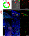
Neonatal myocardium allows for extended persistence of c‐Kit+ cardiac interstitial cells (cCICs). A, Schematic of neonatal injection at P3 and sample collection at 7‐day interval for 28 days. B, Tilescan showing cCICs are retained as patches within left ventricular (LV) wall at 7 days postinjection (dpi; n = 5/5). C, Zoomed‐in view of boxed area in (B) showing cCICs do not colocalize with cardiomyocytes (Desmin, green) at 7 dpi. D, cCICs express TenC at early injection period. E, Tilescan showing cCICs are integrated within LV wall at 14 dpi (n = 6/6). F, Zoomed‐in view of boxed area in (E) showing cCICs share tight junctions (ZO1, green) with resident neighboring host cells at 14 dpi. G, cCICs intercalated among resident cardiomyocytes (Desmin, green) at 14 dpi. H, Tilescan of cCICs persistence at LV apex area at 21 dpi (n = 9/16). I, Zoom‐in of boxed area in (H) showing cCICs spindle morphology and closely localized to neighboring cardiomyocytes (Desmin, green). J, cCICs continue to express TenC at 21 dpi. K, Tilescan showing cCICs persist at LV apex area at 28 dpi (n = 3/3). L, Zoomed‐in view of boxed area in (K). M, cCICs do not colocalize with cardiomyocytes (Desmin, green) at 28 dpi
Figure 4
Figure 4
Engrafted c‐Kit+ cardiac interstitial cells (cCICs) remain active in cell cycle for up to 14 days revealed by fluorescence ubiquitination‐based cell cycle indicators (FUCCI). A, Schematic of FUCCI fluorescence oscillation and cell cycle progression. B, Morphology of FUCCI lentiviral engineered cCICs expressing monomeric Kusabira Orange (mKO; G1 phase) and AzG (S/G2/M phases) fluorescence. BF, bright field. C‐E, Following neonatal (P3) intramyocardial injection, majority cCICs express both mKO and AzG at 7 dpi. Boxed area represented in (D, merged) and (E, mKO and AzG) (n = 3). F‐H, cCICs are still proliferative at 14 dpi indicated by AzG expression (green). Boxed area represented in (g, merged) and (h, mKO and AzG) (n = 3). I‐K, Majority of retained cCICs not proliferative at 21 dpi indicated mKO+ (red) AzG‐ expression (green). Boxed area represented in (J, merged) and (K, mKO and AzG) (n = 3). Scale bar, 50 μm
Figure 5
Figure 5
Neonatal cardiac structural and functional development is not compromised by c‐Kit+ cardiac interstitial cell (cCIC) persistence. A, Masson's Trichrome staining of phosphate‐buffered saline‐injected and cCIC‐injected hearts at 21 and 28 dpi. Small fibrotic area at 28 dpi in the left ventricular apex (arrowhead). B, Immunostaining of myocardium (cTnI) surrounding immediate injection zone (left, *), border zone (middle, *), and remote zone (right), showing structure of myocardium is morphologically normal at 28 dpi. C, Parasternal long‐axis echocardiography at P30, showing injected hearts are comparable to sham operated animals. Left: Sham, uninjected. Right: cCIC‐injected. D, Cardiac physiological functions are comparable between injected and uninjected animals. EF, ejection fraction. FS, fractional shortening. Unpaired Student's t test, two‐tailed (n = 3 hearts for each group). Scale bar, 100 μm
Figure 6
Figure 6
Polyploid DNA content of c‐Kit+ cardiac interstitial cell (cCIC) consistent with extra‐embryonic membrane localization following blastocyst injections. A, cCICs possess tetraploid (4n) DNA content relative to sperm (haploid, 1n) and bone marrow cell (BMC; diploid, 2n) as shown by flow cytometry. B and C, cCICs tetraploidy confirmed by confocal microscopy relative to BMC and sperm. Left, nuclear morphology. Right, quantitation of DAPI intensity (n = 26 for sperms, n = 19 for BMCs, n = 24 for cCICs). D, Cartoon model showing trophectoderm (TE)‐integrated cCICs (red) transitioning into patches in the AM (arrowhead), whereas ICM primarily gives rise to embryo proper (light pink). Scale bar, 10 μm

Similar articles

Cited by

References

    1. Hatzistergos KE, Takeuchi LM, Saur D, et al. cKit+ cardiac progenitors of neural crest origin. Proc Natl Acad Sci. 2015;112:13051‐13056. - PMC - PubMed
    1. Gude NA, Broughton KM, Firouzi F, Sussman MA. Cardiac ageing: extrinsic and intrinsic factors in cellular renewal and senescence. Nat Rev Cardiol. 2018;15:523‐542. - PubMed
    1. Gude NA, Firouzi F, Broughton KM, et al. Cardiac c‐kit biology revealed by inducible transgenesis. Circ Res. 2018;123:57‐72. - PMC - PubMed
    1. Kanisicak O, Khalil H, Ivey MJ, et al. Genetic lineage tracing defines myofibroblast origin and function in the injured heart. Nat Commun. 2016;7:12260. - PMC - PubMed
    1. Yu J, Seldin MM, Fu K, et al. Topological arrangement of cardiac fibroblasts regulates cellular plasticity. Circ Res. 2018;123:73‐85. - PMC - PubMed

Publication types