

Published Erratum
doi: 10.1093/brain/awz409.
Corrigendum
- PMID: 31891369
- PMCID: PMC7089655
- DOI: 10.1093/brain/awz409
Item in Clipboard
Published Erratum
Corrigendum
Brain.
.
No abstract available
Figures
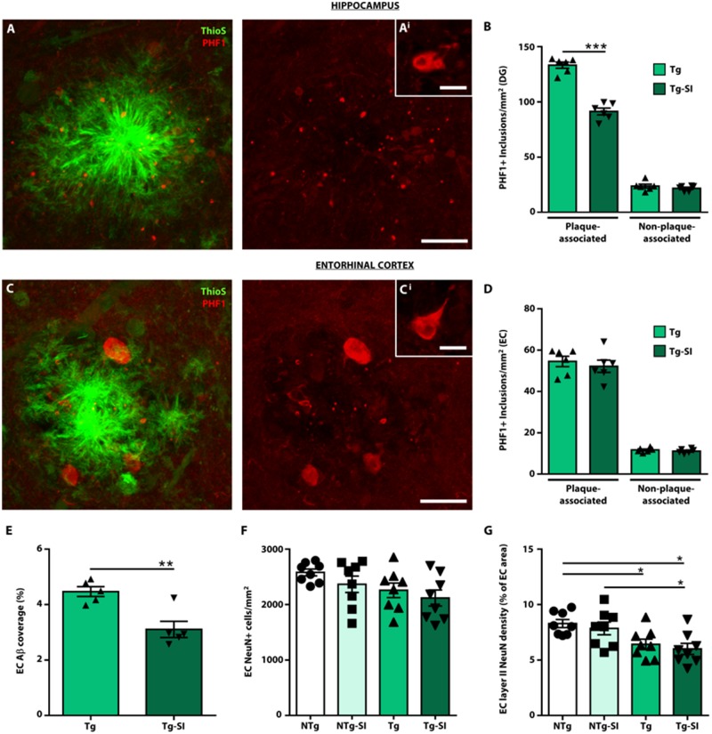
Amyloid-β attenuation decreases tau pathology in a brain region-dependent manner. Tau pathology was assessed in TgF344-AD rats (Tg, Tg-SI) by immunostaining with PHF1 to label hyperphosphorylated tau at S396/404 and Thioflavin-S (Thio-S) to label amyloid-β plaques. (A) PHF1+/Thio-S+ staining in the dentate gyrus (DG) of a Tg rat. Approximately 80–85% of PHF1+ inclusions (red) associate with amyloid-β plaques (green), representing dystrophic neurites. [A(i)] The remainder of the tau inclusions are non-plaque associated, representing pretangle inclusions. (B) PHF1+ inclusions were separated into plaque and non-plaque associated, and quantified by immunohistochemistry (Supplementary Fig. 7) in the dentate gyrus. Amyloid-β attenuation significantly decreased plaque-associated, but not non-plaque associated inclusions. [C and C(i)] PHF1+/Thio-S+ staining in the entorhinal cortex of a Tg rat indicating plaque-associated inclusions and non-plaque associated inclusions. (D) There was no effect of treatment on either plaque-associated or non-plaque associated inclusions in the entorhinal cortex (EC) (E), despite a reduction of amyloid-β plaques in the region. (F) No significant differences were detected in the number of total entorhinal cortical neurons across genotype and treatment (G); however, Tg and Tg-SI rats exhibit a decrease of neurons within layer II of the entorhinal cortex. Scale bars = 20 μm in A and C; 10 μm in A(i) and C(i). n = 6, mean ± SEM, two-sided unpaired t-test, ***P < 0.001.
Erratum for
-
Regional differences in Alzheimer's disease pathology confound behavioural rescue after amyloid-β attenuation.Brain. 2020 Jan 1;143(1):359-373. doi: 10.1093/brain/awz371. Brain. 2020. PMID: 31782760 Free PMC article.
