. 2020 Mar 1;318(3):C463-C475.
doi: 10.1152/ajpcell.00284.2018. Epub 2019 Dec 31.

Endothelial nitric oxide synthase activation is required for heparin receptor effects on vascular smooth muscle cells

Affiliations

Endothelial nitric oxide synthase activation is required for heparin receptor effects on vascular smooth muscle cells

Yaqiu Li et al. Am J Physiol Cell Physiol. .

Abstract

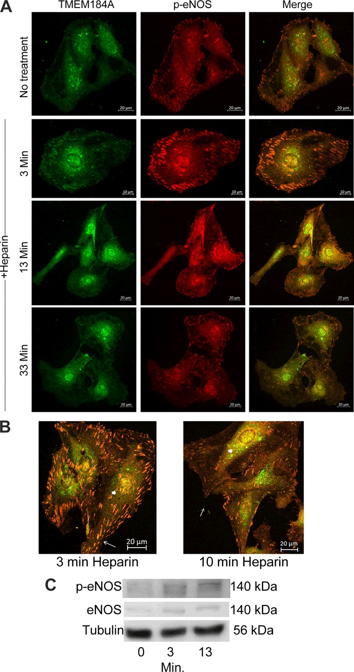
Published studies indicate that TMEM184A is a heparin receptor that interacts with and transduces stimulation from heparin in vascular cells. Previous studies have indicated that heparin increases endothelial nitric oxide synthase (eNOS) activity in bovine endothelial cells. However, the precise mechanism remains unknown. In this study, we investigated the impact of heparin treatment and TMEM184A on eNOS's activation and the role of eNOS in heparin signaling in the cloned A7r5 rat vascular smooth muscle cell line and confirmed results in endothelial cells. We employed a combination of TMEM184A knockdown A7r5 cells along with transient eNOS knockdown and enzyme inhibitor strategies. The results indicate that heparin induces phosphorylation of eNOS. eNOS can be immunoprecipitated with TMEM184A and is internalized to the perinuclear region in a TMEM184A-dependent manner in response to heparin. We also examined how heparin treatment leads to phosphorylation of eNOS and confirmed that TMEM184A and Ca2+ were required to mediate heparin-elicited eNOS phosphorylation. Evidence supporting the involvement of transient receptor potential cation channel subfamily V member 4 with TMEM184A in this eNOS activation process is also presented.

Keywords: eNOS; heparin; vascular smooth muscle.


Conflict of interest statement

No conflicts of interest, financial or otherwise, are declared by the authors.

Figures

Fig. 1.
Fig. 1.
Knockdown (KD) of endothelial nitric oxide synthase (eNOS) eliminates heparin responses in A7r5 cells. A: A7r5 cells transfected with siRNA against eNOS or control siRNA were treated with or without 200 μg/mL heparin for 10 min followed by fixation with ice-cold MeOH. N/T, not treated. Cells were stained for eNOS. Fluorescence intensity of at least 100 cells from 3 separate experiments was analyzed for each condition. ***P < 0.005. Scale bars = 50 μm. B: identical cells were prepared as in A. At least 50 cells per condition in each of 3 individual experiments were analyzed for pERK. ***P < 0.0001. C: eNOS siRNA-transfected A7r5 cells and control siRNA-transfected A7r5 cells were treated with 200 µg/mL heparin for 10 min followed by 1.5 µg/mL PDGF stimulation. Cells were stained for pELK. ***P < 0.0001, heparin/PDGF compared with PDGF. The graph represents data from 3 independent experiments with at least 50 cells analyzed per condition in each experiment.
Fig. 2.
Fig. 2.
Endothelial nitric oxide synthase (eNOS) colocalizes with transmembrane protein 184A (TMEM184A) before and after heparin treatment. A: A7r5 cells were incubated with 200 μg/mL heparin for the indicated times and fixed and stained for p-eNOS (red) and TMEM184A (green). Images are representative of 2 separate experiments. Scale bars: no treatment = 20 μm; 3 min = 10 µm; 13 and 33 min = 30 µm. B: A7r5s treated as in A taken at a higher magnification show colocalization between p-eNOS and TMEM184A at focal adhesions (arrows) and perinuclear regions (arrowheads). Scale bars = 20 µm. C: A7r5 cells were not treated, or treated with heparin for 3 or 13 min, harvested as described, placed into sample buffer, and separated on SDS polyacrylamide gels. Western blots were developed using the CST rabbit eNOS antibody or p-Ser 1177 eNOS antibody. The blots are representative of at least 3 separate experiments.
Fig. 3.
Fig. 3.
Transmembrane protein 184A (TMEM184A) knockdown (KD) A7r5 cells do not show heparin-induced increase in p-Ser 1177 endothelial nitric oxide synthase (eNOS). A: A7r5 cells and stable TMEM184A A7r5 knockdown cells were treated with 200 μg/mL heparin for the indicated times followed by fixation and staining for p-eNOS. Images are representative of 3 independent experiments. Scale bars = 50 μm. B: A7r5 cells and TMEM184A stable knockdown cells were treated with heparin as in A. At least 50 cells per condition from 3 separate experiments were evaluated for p-eNOS. ***P < 0.0001, for heparin vs. no heparin.
Fig. 4.
Fig. 4.
Transmembrane protein 184A (TMEM184A) interacts with endothelial nitric oxide synthase (eNOS) and αV integrin. A: membrane protein samples of bovine aortic endothelial cells (BAOECs) and A7r5 lysates were incubated with the NOS3 antibody (sc-8331) and precipitated using EZview affinity beads, or the lysates were incubated with beads without antibodies (B) and Western blots were developed using TMEM184A (internal domain) antibody. IB, immunoblot. The BAOEC blot is representative of 2 separate experiments. The BAOEC blot shown also contained a sample of whole cell lysate. B: A7r5 cells were harvested for immunoprecipitation (IP). Cell samples were incubated with TMEM184A (NTD, NH2-terminal domain) antibody and precipitated using EZview affinity beads or beads and cell lysates without antibody (B) and blots developed using integrin αV antibody. The blot is representative of 2 separate experiments.
Fig. 5.
Fig. 5.
EGTA abolished heparin-induced activation of endothelial nitric oxide synthase (eNOS). A: A7r5 cells preincubated for 30 min with or without EGTA were treated for 13 min with or without 200 μg/mL heparin followed by fixation with ice-cold MeOH and staining for p-eNOS. Images are representative of 3 independent experiments. Scale bars = 50 μm. B: A7r5 cells were treated with heparin and EGTA as in A. In each experiment, at least 50 cells per condition were examined for p-eNOS. ***P < 0.0001, heparin treated vs. without heparin. C: A7r5 cells preincubated for 30 min with or without EGTA were then treated with or without heparin for 3 or 13 min and analyzed by Western blotting with antibodies against p-Ser 1177 eNOS and actin (produced in mouse) and developed with Cy3 tagged anti-rabbit secondary antibodies and Alexa 488 tagged anti-mouse secondary antibodies. D: less confluent cells treated as in C were analyzed by Western blotting with antibodies against pERK and tubulin with fluorescent secondary antibodies.
Fig. 6.
Fig. 6.
KN-93 treatment decreased heparin-induced activation of endothelial nitric oxide synthase (eNOS). A: A7r5 cells preincubated for 30 min with or without KN-93 were treated with or without 200 μg/mL heparin for 13 min followed by fixation with ice-cold MeOH and staining for p-Ser 1177 eNOS. Images are representative of 3 independent experiments. Scale bars = 50 μm. B: A7r5 cells were treated with heparin and KN-93 as in A. At least 50 cells per condition were examined for p-eNOS in each repeat. ***P < 0.0001, heparin vs. without heparin and KN-93 heparin vs. control heparin.
Fig. 7.
Fig. 7.
Transient receptor potential cation channel subfamily V member 4 (TRPV4) inhibitors decreased heparin-induced activation of endothelial nitric oxide synthase (eNOS). A and B: A7r5 cells preincubated for 30 min with or without RN1734 were treated with or without 200 μg/mL heparin followed by fixation and staining for p-eNOS. Images are representative of 3 independent experiments. Scale bars = 50 μm. C and D: A7r5 cells were treated with GSK and heparin as in A and B. At least 50 cells per condition were examined for p-eNOS in each repeat. **P < 0.0001, heparin vs. without heparin. E: A7r5 cells treated as in A and B were harvested for Western blotting, stained with antibodies against p-eNOS, total eNOS (5880), and tubulin, and blots were developed with secondary fluorescently tagged antibodies as in Fig. 5. F: less confluent A7r5 cells treated as in A and B were harvested for Western blotting; stained with antibodies against p-eNOS, pERK, and tubulin, and developed with fluorescently tagged antibodies as in Fig. 5.
Fig. 8.
Fig. 8.
Endothelial nitric oxide synthase (eNOS) in bovine aortic endothelial cells (BAOECs) also gains p-Ser 1177 in response to heparin treatment. A: BAOECs were left untreated (C) or treated with either VEGF at 100 ng/mL (V) or heparin at 200 μg/mL (H) for 5 min. Cells were harvested for eNOS Western blotting for p-Ser 1177 eNOS and tubulin as a loading control. B: BAOECs were pretreated with EGTA as in Fig. 5 and then treated with heparin or not. Cells were harvested and analyzed by Western blotting for p-Ser 1177 eNOS, total eNOS (5880 mouse antibody) and tubulin. Blots were developed with fluorescently tagged antibodies. C: BAOECs were treated with RN1734 as in Fig. 7 and analyzed as in B.
Fig. 9.
Fig. 9.
A: bovine aortic endothelial cells (BAOECs) were stained for αV integrin (red) and total endothelial nitric oxide synthase (eNOS; 5880) (green). B: BAOECs were grown on coverslips, treated with 200 μg/mL heparin for 3 or 10 min or left untreated, (fixed as noted in experimental procedures) and stained for p-Ser 1177 eNOS (green) and αV integrin (white). C: BAOECs were treated with heparin and stained with αV integrin (white), transmembrane protein 184A (TMEM184A; NH2-terminal domain; red), and total eNOS (5880, green). Scale bars = 20 µm. Zoomed sections of merge-3 images were altered to remove the αV integrin channel for clarity of the limited colocalization of TMEM184A and total eNOS. Scale bars on zoomed images are 10 µm.
Fig. 10.
Fig. 10.
Model for the signal steps between transmembrane protein 184A (TMEM184A) and endothelial nitric oxide synthase (eNOS) phosphorylation. This model indicates likely interactions between TMEM and transient receptor potential cation channel subfamily V member 4 (TRPV4). The mechanism for this interaction is unknown and represented by dashes. The remaining connections shown have been established by others, and our studies indicate roles for these in the response to heparin.

Similar articles

Cited by

References

    1. Akimoto S, Mitsumata M, Sasaguri T, Yoshida Y. Laminar shear stress inhibits vascular endothelial cell proliferation by inducing cyclin-dependent kinase inhibitor p21(Sdi1/Cip1/Waf1). Circ Res 86: 185–190, 2000. doi:10.1161/01.RES.86.2.185. - DOI - PubMed
    1. Arias-Díaz J, Vara E, Torres-Melero J, García C, Hernández J, Balibrea JL. Local production of oxygen free radicals and nitric oxide in rat diaphragm during sepsis: effects of pentoxifylline and somatostatin. Eur J Surg 163: 619–625, 1997. - PubMed
    1. Arredondo Zamarripa D, Noguez Imm R, Bautista Cortés AM, Vázquez Ruíz O, Bernardini M, Fiorio Pla A, Gkika D, Prevarskaya N, López-Casillas F, Liedtke W, Clapp C, Thébault S. Dual contribution of TRPV4 antagonism in the regulatory effect of vasoinhibins on blood-retinal barrier permeability: diabetic milieu makes a difference. Sci Rep 7: 13094, 2017. [Erratum in Sci Rep 8: 9652, 2018.] doi:10.1038/s41598-017-13621-8. - DOI - PMC - PubMed
    1. Ballut L, Sapay N, Chautard E, Imberty A, Ricard-Blum S. Mapping of heparin/heparan sulfate binding sites on αvβ3 integrin by molecular docking. J Mol Recognit 26: 76–85, 2013. doi:10.1002/jmr.2250. - DOI - PubMed
    1. Baratchi S, Knoerzer M, Khoshmanesh K, Mitchell A, McIntyre P. Shear stress regulates TRPV4 channel clustering and translocation from adherens junctions to the basal membrane. Sci Rep 7: 15942, 2017. doi:10.1038/s41598-017-16276-7. - DOI - PMC - PubMed

Publication types

MeSH terms

LinkOut - more resources