. 2020 Mar 1:209:120581.
doi: 10.1016/j.talanta.2019.120581. Epub 2019 Nov 23.

Pixelated colorimetric nucleic acid assay

Affiliations

Pixelated colorimetric nucleic acid assay

Hakan Berk Aydın et al. Talanta. .

Abstract

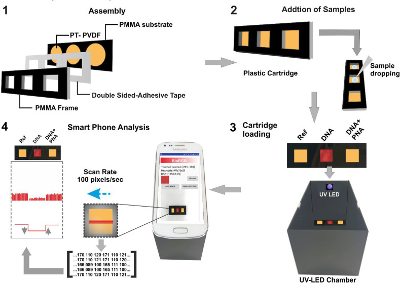
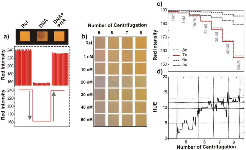
Conjugated polyelectrolytes (CPEs) have been widely used as reporters in colorimetric assays targeting nucleic acids. CPEs provide naked eye detection possibility by their superior optical properties however, as concentration of target analytes decrease, trace amounts of nucleic acid typically yield colorimetric responses that are not readily perceivable by naked eye. Herein, we report a pixelated analysis approach for correlating colorimetric responses of CPE with nucleic acid concentrations down to 1 nM, in plasma samples, utilizing a smart phone with an algorithm that can perform analytical testing and data processing. The detection strategy employed relies on conformational transitions between single stranded nucleic acid-cationic CPE duplexes and double stranded nucleic acid-CPE triplexes that yield distinct colorimetric responses for enabling naked eye detection of nucleic acids. Cationic poly[N,N,N-triethyl-3-((4-methylthiophen-3-yl)oxy)propan-1-aminium bromide] is utilized as the CPE reporter deposited on a polyvinylidene fluoride (PVDF) membrane for nucleic acid assay. A smart phone application is developed to capture and digitize the colorimetric response of the individual pixels of the digital images of CPE on the PVDF membrane, followed by an analysis using the algorithm. The proposed pixelated approach enables precise quantification of nucleic acid assay concentrations, thereby eliminating the margin of error involved in conventional methodologies adopted for interpretation of colorimetric responses, for instance, RGB analysis. The obtained results illustrate that a ubiquitous smart phone could be utilized for point of care colorimetric nucleic acids assays in complex matrices without requiring sophisticated software or instrumentation.

Keywords: Conjugated polyelectrolyte; Diagnostic tool; Nucleic acid assay; Paper-based sensor; Pixelated analysis; Point-of-care.


Conflict of interest statement

The authors declare no conflict of interest.

Figures

Image 1
Graphical abstract
Scheme 1
Scheme 1
Schematic illustration of cartridge and smart phone-assisted nucleic acid assay. Step 1 illustrates the assembly of plastic cartridge consisting of three layers; Step 2 shows the transfer of HBV DNA sample to the cartridge and Step 3 shows the mounting cartridge on the UV-LED chamber that serves as an accessory for mounting the smart phone. Step 4 illustrates the smart-phone assisted BioRGB analysis yielding bar graphs that represents the pixelated (and averaged) RED intensities of the three sample spots.
Fig. 1
Fig. 1
Scanning and analysis of samples spots on cartridge by the smart phone application; a) the typical scan process generates matrix consisting of RGB color codes that is transformed to the bar graphs for display at the user interface of the application. b) Microstain generated by artificial distortion of 4 × 4 pixels, 25 times magnified image of the microstain and its corresponding red intensity profile are shown in (c), d) Evaluation of droplet pattern recognition performance of application: the snapshot of droplet pattern (edges were intentionally marked by white lines for sake of better visualization) and (e) corresponding red, green and blue intensity profiles of droplet pattern shown in d. (For interpretation of the references to color in this figure legend, the reader is referred to the Web version of this article.)
Fig. 2
Fig. 2
a) Red intensity profiles of reference (no HBV DNA), control HBV DNA (single stranded) and sample HBV DNA-PNA (hybridization). b) Images of PT on PVDF membrane with increasing centrifugation of PT from five to eight times (from left to right); the change in the color of PT coated PVDF with increasing HBV DNA concentration from 0 to 50 nM (from top to bottom). c) and d) red intensity and hue profiles obtained from the images shown Fig. 2b. (For interpretation of the references to color in this figure legend, the reader is referred to the Web version of this article.)
Fig. 3
Fig. 3
a) The red intensity profile for 1 nM, 10 nM, 100 nM and 1000 nM of HBV DNA and calculation of Δ RED (b), the algebraic transformation of color matrix. (c) residual plot showing scattering of less than 1% for Δ RED and d) the calibration curve for HBV DNA detection. (For interpretation of the references to color in this figure legend, the reader is referred to the Web version of this article.)
Fig. 4
Fig. 4
a) Digital images of cartridges utilized for 1 nM, 10 nM, 100 nM and 1000 nM of HBV DNA spiked in plasma, b) corresponding BioRGB red intensity profiles and c) scattering intensities. (For interpretation of the references to color in this figure legend, the reader is referred to the Web version of this article.)

References

    1. Mehrotra P. Biosensors and their applications - a review. J. Oral Biol. Craniofac. Res. 2016;6(2):153–159. - PMC - PubMed
    1. Justino C.I.L., Duarte A.C., Rocha-Santos T.A.P. Critical overview on the application of sensors and biosensors for clinical analysis. Trac. Trends Anal. Chem. 2016;85:36–60. - PMC - PubMed
    1. Lifson M.A., Ozen M.O., Inci F., Wang S., Inan H., Baday M., Henrich T.J., Demirci U. Advances in biosensing strategies for HIV-1 detection, diagnosis, and therapeutic monitoring. Adv. Drug Deliv. Rev. 2016;103:90–104. - PMC - PubMed
    1. Baeumner A.J., Schlesinger N.A., Slutzki N.S., Romano J., Lee E.M., Montagna R.A. Biosensor for dengue virus Detection: sensitive, rapid, and serotype specific. Anal. Chem. 2002;74(6):1442–1448. - PubMed
    1. Choi J.R., Tang R., Wang S., Wan Abas W.A., Pingguan-Murphy B., Xu F. Paper-based sample-to-answer molecular diagnostic platform for point-of-care diagnostics. Biosens. Bioelectron. 2015;74:427–439. - PubMed