

Extraction of Hydroxyapatite Nanostructures from Marine Wastes for the Fabrication of Biopolymer-Based Porous Scaffolds
- PMID: 31892123
- PMCID: PMC7024202
- DOI: 10.3390/md18010026
Extraction of Hydroxyapatite Nanostructures from Marine Wastes for the Fabrication of Biopolymer-Based Porous Scaffolds
Abstract
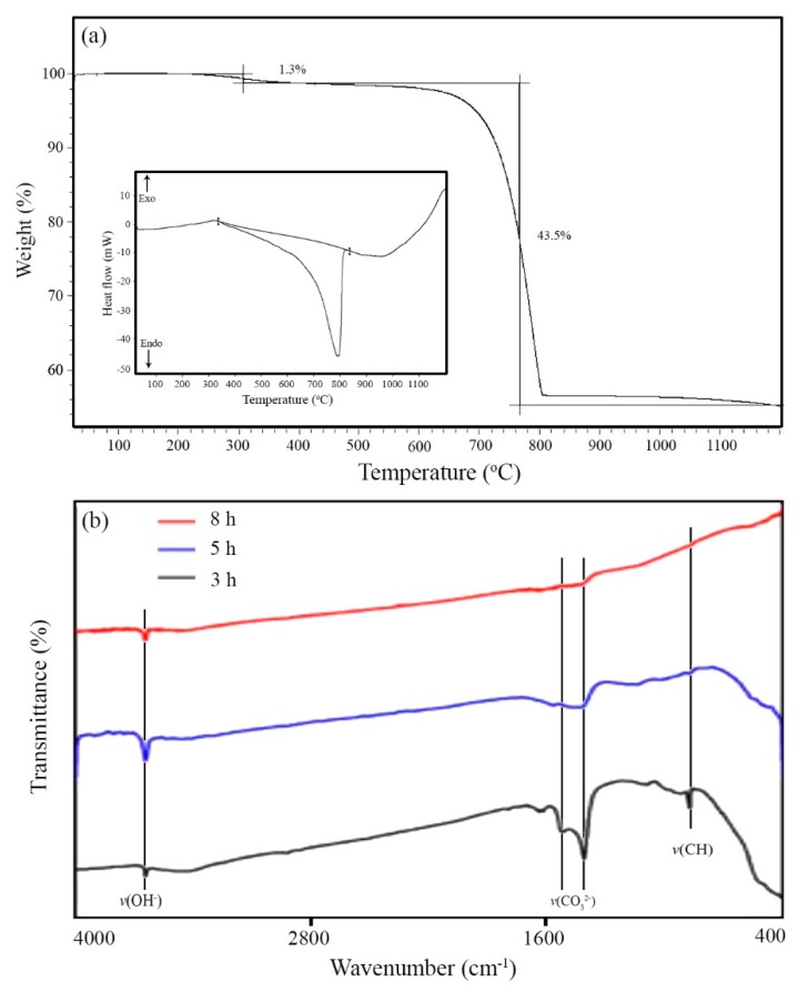
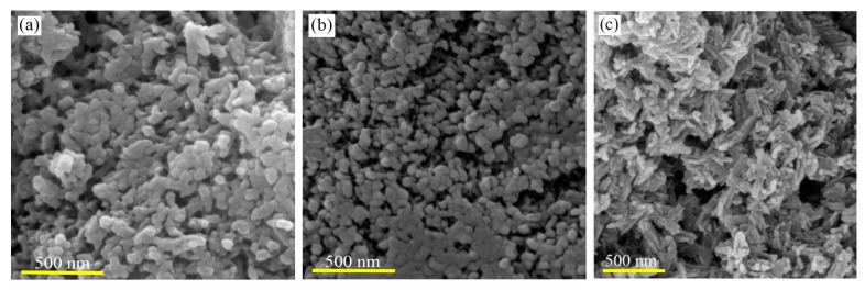
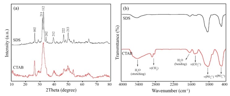
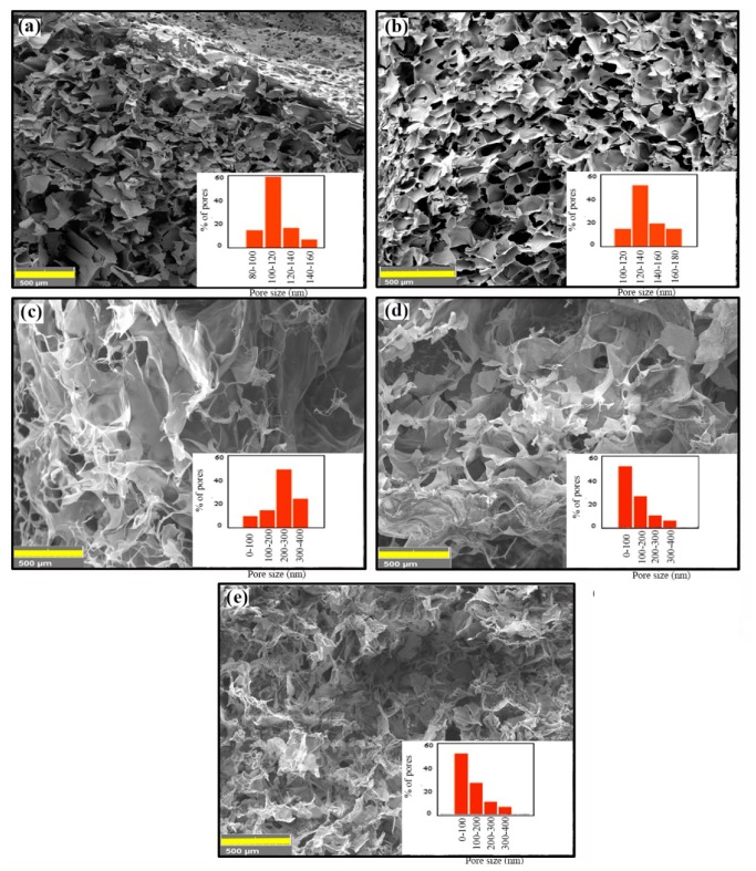
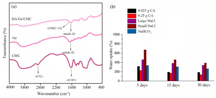
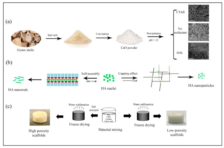
Three-dimensional porous nanocomposites consisting of gelatin-carboxymethylcellulose (CMC) cross-linked by carboxylic acids biopolymers and monophasic hydroxyapatite (HA) nanostructures were fabricated by lyophilization, for soft-bone-tissue engineering. The bioactive ceramic nanostructures were prepared by a novel wet-chemical and low-temperature procedure from marine wastes containing calcium carbonates. The effect of surface-active molecules, including sodium dodecyl sulfate (SDS) and hexadecyltrimethylammonium bromide (CTAB), on the morphology of HA nanostructures is shown. It is demonstrated that highly bioactive and monophasic HA nanorods with an aspect ratio > 10 can be synthesized in the presence of SDS. In vitro studies on the bioactive biopolymer composite scaffolds with varying pore sizes, from 100 to 300 μm, determine the capacity of the developed procedure to convert marine wastes to profitable composites for tissue engineering.
Keywords: biopolymer; cytotoxicity; marine waste; tissue engineering.
Conflict of interest statement
The authors declare no potential conflicts of interest with respect to the research, authorship, and/or publication of this article.
Figures

References
-
- Ehrlich H., Douglas T., Scharnweber D., Hanke T., Born R., Bierbaum S., Worch H. Hydroxyapatite crystal growth on modified collagen I-templates in a model dual membrane diffusion system. Z. Anorg. Allg. Chem. 2005;631:1825–1830. doi: 10.1002/zaac.200500195. - DOI
-
- Ehrlich H. Chitin and collagen as universal and alternative templates in biomineralization. Int. Geol. Rev. 2010;52:661–699. doi: 10.1080/00206811003679521. - DOI
MeSH terms
Substances
Grants and funding
LinkOut - more resources
Full Text Sources
