. 2019 Dec 27;12(1):82.
doi: 10.3390/nu12010082.

Pterostilbene Prevents Early Diabetic Retinopathy Alterations in a Rabbit Experimental Model

Affiliations

Pterostilbene Prevents Early Diabetic Retinopathy Alterations in a Rabbit Experimental Model

Iván Millán et al. Nutrients. .

Abstract

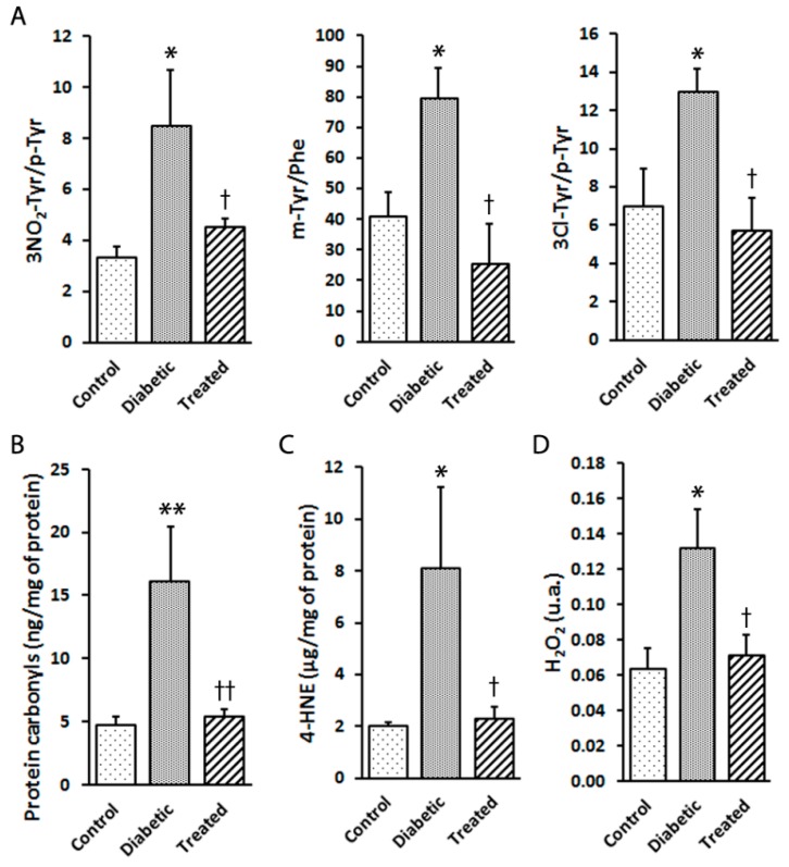
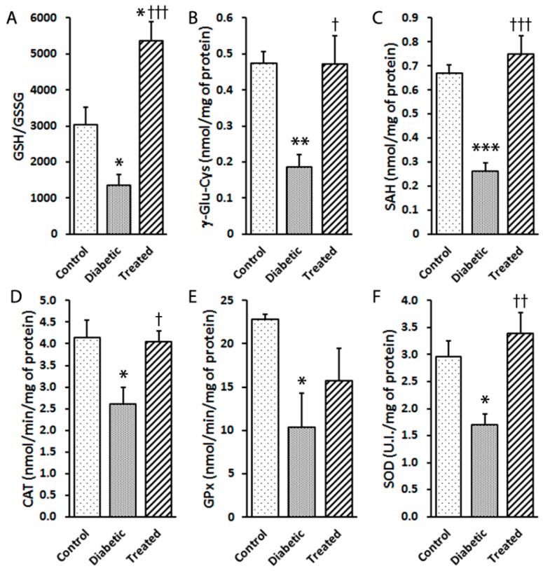
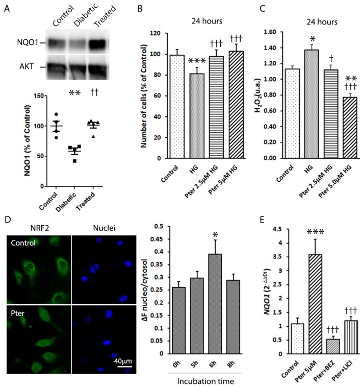
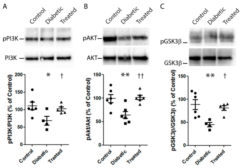
Oxidative stress generated by diabetes plays a key role in the development of diabetic retinopathy (DR), a common diabetic complication. DR remains asymptomatic until it reaches advanced stages, which complicate its treatment. Although it is known that good metabolic control is essential for preventing DR, knowledge of the disease is incomplete and an effective treatment with no side effects is lacking. Pterostilbene (Pter), a natural stilbene with good antioxidant activity, has proved to beneficially affect different pathologies, including diabetes. Therefore, our study aimed to analyse the protective and/or therapeutic capacity of Pter against oxidant damage by characterising early retinal alterations induced by hyperglycaemia, and its possible mechanism of action in a rabbit model of type 1 diabetes mellitus. Pter reduced lipid and protein oxidative damage, and recovered redox status and the main activities of antioxidant enzymes. Moreover, the redox regulation by Pter was associated with activation of the PI3K/AKT/GSK3β/NRF2 pathway. Our results show that Pter is a powerful protective agent that may delay early DR development.

Keywords: diabetic retinopathy; oxidative stress; polyphenol; pterostilbene; retinal damage.


Conflict of interest statement

The authors declare no conflict of interest. The funders played no role in: the study design; collections; analyses; data interpretations; writing the manuscript; deciding to publish the results.

Figures

Figure 1
Figure 1
Chemical structures and plasma levels of Pter after administering Pter phosphate disodium salt. The pharmaceutical form of disodium salt of Pter phosphate was used to facilitate its daily subcutaneous administration in vivo. (A) Chemical structures of Pter and Pter phosphate disodium salt. (B) A subcutaneous injection of Pter phosphate disodium salt (Pter 50 mg/kg) was administered dissolved in water (25 mg/mL). Blood was collected at different times and Pter was quantified in plasma by the UPLC-MS/MS analysis. The results are the means ± S.D. of four different rabbits.
Figure 2
Figure 2
Type 1 diabetes mellitus was induced in rabbits by alloxan injection and rabbits were treated daily with Pter phosphate disodium salt (50 mg/kg of Pter) for six weeks (Treated). (A) Weight (BW) and (B) blood glucose (BG) levels were evaluated weekly. (C) Retinal apoptotic cell death was examined and quantified by TUNEL staining (black bar = 50 µm) six weeks after diabetes induction. Black arrows mark apoptotic cells. Data are presented as mean ± S.E.M. (at least n = 5 in each group). An ANOVA, followed by Tukey’s post hoc test, was used to assess significant differences between experimental conditions. * p < 0.05; ** p < 0.01; *** p < 0.001 versus the control group. † p < 0.05; ††† p < 0.001 versus the diabetic group.
Figure 3
Figure 3
Effect of pterostilbene treatment on the oxidation of proteins and lipids, as well as hydrogen peroxide production, in the rabbit retinas. Retinal samples from the control, diabetic and diabetic and Pter-treated rabbits were obtained six weeks after inducing diabetes. (A) The 3NO2-Tyr/p-Tyr, m-Tyr/Phe and 3Cl-Tyr/p-Tyr ratios were quantified by UPLC-MS/MS. (B) Carbonylation was measured in retinal homogenates by ELISA. (C) Lipid peroxidation was determined by ELISA. (D) Hydrogen peroxide was established in retina homogenates by spectrofluorometry. Data are presented as mean ± SEM (at least n = 4 in each group). Data were analysed by a one-way ANOVA followed by Newman-Keuls test. * p < 0.05; ** p < 0.01 versus the control group. † p < 0.05; †† p < 0.01 versus the diabetic group.
Figure 4
Figure 4
Effect of pterostilbene on antioxidant machinery activation in diabetic rabbits. Type 1 diabetes mellitus and Pter treatment were performed as described in Figure 2. The analyses of retinal (A) GSH/GSSG, (B) γ-Glu-Cys and (C) SHA were performed by UPLC-MS/MS. The enzymatic activities of (D) catalase (CAT), (E) glutathione peroxidase (GPx) and (F) superoxide dismutase (SOD) were determined by spectrophotometry. Data are presented as mean ± SEM (at least n = 4 in each group). Statistical analyses were performed by a one-way ANOVA followed by Tukey’s test. * p < 0.05; ** p < 0.01; *** p < 0.001 versus the control group. † p < 0.05; †† p < 0.01; ††† p < 0.001 versus the diabetic group.
Figure 5
Figure 5
Effect of pterostilbene on NRF2 activation. (A) A Western blot was used to detect the expressions of NQO1 in retinas. Data are presented as mean ± SEM (at least n = 4 in each group). A one-way ANOVA and a Newman-Keuls multiple comparison test were used. ** p < 0.01; versus the control group; †† p < 0.01 versus the diabetic group. (B) HREC were treated for 24 h with Pter (2.5 μM and 5 μM) at high (30 mM) glucose concentrations. Statistical analyses were performed by a one-way ANOVA, followed by Tukey’s test. *** p < 0.001 versus the control group. ††† p < 0.001 versus the high glucose group. (C) Hydrogen peroxide was determined in HREC medium after 24 h of incubation with Pter (2.5 μM and 5 μM) at high (30mM) glucose concentrations. An ANOVA followed by Tukey’s post hoc test, was used to assess significant differences between the experimental conditions * p < 0.05; ** p < 0.01; versus the control group. † p < 0.05; ††† p < 0.001 versus the high glucose group. (D) NRF2 translocation was detected by confocal microscopy in HREC. Cells were incubated with Pter 5 μM and fixed with 4% paraformaldehyde at 0, 5, 6 and 8 h. NRF2 was detected by immunocytochemistry and NRF2 translocation was evaluated by the ImageJ software. A representative image after 6 h of incubation with Pter. Data were analysed by a one-way ANOVA, followed by Tukey’s test. * p < 0.05 versus the control group (0 h) (E) HREC were incubated in the presence of DMSO (control), Pter 5 μM, Pter (5 uM) + BEZ235 (10 nM), and Pter (5 uM) + LiCl (20 mM) for 24 h. The RNA levels of NQO1 were determined by qRT-PCR normalised to the β-Actin mRNA levels. Data are presented as mean ± SEM (at least n = 3 in each group). Differences among groups were assessed by a one-way ANOVA, followed by Tukey’s test. *** p < 0.001 versus the control group. ††† p < 0.001 versus the Pter group.
Figure 6
Figure 6
Effect of pterostilbene on the PI3K/AKT/GSK3β/NRF2 pathway. Type 1 diabetes mellitus and Pter treatment were performed as described in Figure 2. A Western blot was used to detect the expressions of pPI3K/PI3K (A), pAKT/AKT (B), pGSK3β/GSK3β (C). Data are presented as mean ± SEM (at least n = 4 in each group). An ANOVA, followed by a Newman–Keuls post hoc test, was used to assess any significant differences between the experimental conditions. * p < 0.05; ** p < 0.01 versus the control group. † p < 0.05; †† p < 0.01 versus the diabetic group.

References

    1. WHO . Global Report on Diabetes. World Health Organization; Geneva, Switzerland: 2016.
    1. Maric-Bilkan C. Sex differences in micro- and macro-vascular complications of diabetes mellitus. Clin. Sci. (Lond.) 2017;131:833–846. doi: 10.1042/CS20160998. - DOI - PubMed
    1. Rossino M.G., Casini G. Nutraceuticals for the Treatment of Diabetic Retinopathy. Nutrients. 2019;11:771. doi: 10.3390/nu11040771. - DOI - PMC - PubMed
    1. Wangsa-Wirawan N.D., Linsenmeier R.A. Retinal oxygen: Fundamental and clinical aspects. Arch. Ophthalmol. 2003;121:547–557. doi: 10.1001/archopht.121.4.547. - DOI - PubMed
    1. Kisic B., Miric D., Zoric L. Free Radical Biology of Eye Diseases. In: Laher I., editor. Systems Biology of Free Radicals and Antioxidants. Springer; Berlin/Heidelberg, Germany: 2014. pp. 3599–3623.

MeSH terms