. 2019 Dec 27;21(1):214.
doi: 10.3390/ijms21010214.

Design, Synthesis and Molecular Modeling Study of Conjugates of ADP and Morpholino Nucleosides as A Novel Class of Inhibitors of PARP-1, PARP-2 and PARP-3

Affiliations

Design, Synthesis and Molecular Modeling Study of Conjugates of ADP and Morpholino Nucleosides as A Novel Class of Inhibitors of PARP-1, PARP-2 and PARP-3

Yuliya V Sherstyuk et al. Int J Mol Sci. .

Abstract

We report on the design, synthesis and molecular modeling study of conjugates of adenosine diphosphate (ADP) and morpholino nucleosides as potential selective inhibitors of poly(ADP-ribose)polymerases-1, 2 and 3. Sixteen dinucleoside pyrophosphates containing natural heterocyclic bases as well as 5-haloganeted pyrimidines, and mimicking a main substrate of these enzymes, nicotinamide adenine dinucleotide (NAD+)-molecule, have been synthesized in a high yield. Morpholino nucleosides have been tethered to the β-phosphate of ADP via a phosphoester or phosphoramide bond. Screening of the inhibiting properties of these derivatives on the autopoly(ADP-ribosyl)ation of PARP-1 and PARP-2 has shown that the effect depends upon the type of nucleobase as well as on the linkage between ADP and morpholino nucleoside. The 5-iodination of uracil and the introduction of the P-N bond in NAD+-mimetics have shown to increase inhibition properties. Structural modeling suggested that the P-N bond can stabilize the pyrophosphate group in active conformation due to the formation of an intramolecular hydrogen bond. The most active NAD+ analog against PARP-1 contained 5-iodouracil 2'-aminomethylmorpholino nucleoside with IC50 126 ± 6 μM, while in the case of PARP-2 it was adenine 2'-aminomethylmorpholino nucleoside (IC50 63 ± 10 μM). In silico analysis revealed that thymine and uracil-based NAD+ analogs were recognized as the NAD+-analog that targets the nicotinamide binding site. On the contrary, the adenine 2'-aminomethylmorpholino nucleoside-based NAD+ analogs were predicted to identify as PAR-analogs that target the acceptor binding site of PARP-2, representing a novel molecular mechanism for selective PARP inhibition. This discovery opens a new avenue for the rational design of PARP-1/2 specific inhibitors.

Keywords: DNA repair; NAD+ analogs; PARP; molecular modeling; morpholino nucleosides.


Conflict of interest statement

The authors declare no conflict of interest.

Figures

Figure 1
Figure 1
The structure of the beta-oxidized nicotinamide adenine dinucleotide (NAD+)-molecule.
Figure 2
Figure 2
Morpholino nucleoside adenosine dinucleotides (MorXppA).
Scheme 1
Scheme 1
Synthesis of the conjugates of adenosine diphosphate (ADP) with 2′-hydroxymethylmorpholino nucleosides. Reagents and conditions: (a) NaIO4, EtOH/H2O, 15 min; (NH4)2B4O7·4H2O, Et3N, 1.5 h; NaBH3CN, 40 min; trifluoroacetic acid (TFA), pH 3–4, 1 h; TrCl, Et3N, dimethylformamide (DMF), 3 h; yield 60%–70%; (b) POCl3, Py, –15 °C, 15 min; 1 M triethylammonium bicarbonate (TEAB), yield 75%–90%; (c) Ph3P/(PyS)2, MeIm, 1,3-dimethyl-2-imidazolidinone (DMI); n-Bu3NH+ salt of AMP; conc. aq. NH3 for compounds 4A,G,C; 80% aq. AcOH (v/v); yield 70%–80%.
Scheme 2
Scheme 2
Synthesis of the conjugates of ADP with 2′-aminomethylmorpholino nucleosides. Reagents and conditions: (a) Ph3P, Im, DCE, I2, 0 °C → rt, 5 h; (b) NaN3, DMF, 12 h; (c) H2, 10% Pd/C, MeOH; (d) Ph3P, CBr4, DMI; (e) Ph3P (2 eq), Py; conc. aq. NH3; (f) ADP n-Bu3N salt, Ph3P, (PyS)2, MeIm, DMI; conc. aq. NH3 for compounds 10A,G,C; 80% aq. AcOH (v/v).
Figure 3
Figure 3
Graphs of Km (left) and Vmax (right) values versus inhibitor concentration. 3-AB, a commercially-available PARP-1 inhibitor [75], was used as a positive control.
Figure 4
Figure 4
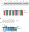
Basic activity of PARP-3 in DNA (A) and protein ADP-ribosylation (B) and the influence of the inhibitors on these reactions. (A) Activity of PARP-3 on [32P]-labeled one-window gapped DNA substrate in the absence (lanes 1–4) or presence of inhibitors in the different concentrations (lanes 5–32) on the upper panel. The reactions were performed using increasing concentration of NAD+. Lane c corresponds to initial electrophoretic mobility of the DNA substrate. The chart on the bottom panel is reflected of the reaction yield of the ADP-ribosylated DNA in percentage terms. (B) Activity of PARP-3 on gap1 DNA substrate in the absence (lanes 1–3) or presence of inhibitors in the different concentrations (lanes 4–18) on the upper panel. The reactions were performed using increasing concentration of NAD+ in the presence of [32P]-labeled NAD+. Lane c corresponds to reaction mixture without PARP-3. The chart on the bottom panel is reflected of the reaction yield of the ADP-ribosylated PARP-3 in the presence of inhibitor normalized on the yield of the autoribosylation of PARP-3 in percentage terms. The ticks on the chart mark the bars with the 0.5 mM of inhibitor in the experiment.
Figure 5
Figure 5
Structural model of the PARP-2 catalytic domain in complex with NAD+ at the donor binding site and ADP fragment at the acceptor binding site. The molecular surface illustrates ADP binding subsites of acceptor substrate (gray color), nicotinamide riboside fragment of donor NAD+ substrate (orange color) and the ADP fragment of donor NAD+ substrate (blue color). HD domain is not shown for simplicity. Substrates are shown in green color.
Figure 6
Figure 6
Predicted binding poses of 11IU (A) and 5-I-Urd (B) bound to the NA binding site of the PARP-2 catalytic domain. Hydrogen bonds are depicted as green dashed lines. Small molecules are shown in green color.
Figure 7
Figure 7
Predicted binding mode of 10IU with the donor binding site of the PARP-2 catalytic domain. (A) The close-view of the donor binding site. (B) Structural alignment of the binding poses of 10IU (green color) with NAD+ (cyan color) and 5-I-Urd (black color). (C) Representative conformations of 10IU from the molecular dynamics trajectory, the conformation with the intramolecular hydrogen bond with the non-bridging α-phosphate oxygen of ADP (bottom) and the conformation with the hydrogen bond with carbonyl oxygen of the heterocyclic base of the modified nucleoside (top) are shown. The molecular surface of the binding site is shown. Hydrogen bonds are depicted as green dashed lines.
Figure 8
Figure 8
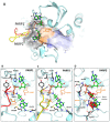
Predicted binding pose of 10A with the acceptor binding site of the PARP-1/2 catalytic domain. (A) The structural alignment of PARP-1 and PARP-2. Variable loops are indicated in red and yellow colors for PARP-1 and PARP-2, respectively. Detailed view of 10A interaction with PARP-1 (B) and PARP-2 (C) is shown. (D) PARP-3 acceptor binding site with superimposed binding pose of 10A from PARP-2/10A complex. Steric clashes are shown with red disks. Unsatisfied hydrogen bond donor and acceptor atoms of the PARP-3 acceptor binding site hindered by ligand are shown as spheres. Hydrogen bonds are depicted as dashed lines. HD domain is not shown for simplicity.

Similar articles

Cited by

References

    1. Amé J.C., Spenlehauer C., de Murcia G. The PARP superfamily. Bioessays. 2004;26:882–893. doi: 10.1002/bies.20085. - DOI - PubMed
    1. Gupte R., Liu Z., Kraus W.L. PARPs and ADP-ribosylation: Recent advances linking molecular functions to biological outcomes. Genes Dev. 2017;31:101–126. doi: 10.1101/gad.291518.116. - DOI - PMC - PubMed
    1. Vyas S., Matic I., Uchima L., Rood J., Zaja R., Hay R.T., Ahel I., Chang P. Family-wide analysis of poly(ADP-ribose) polymerase activity. Nat. Commun. 2014;5:4426. doi: 10.1038/ncomms5426. - DOI - PMC - PubMed
    1. Lüscher B., Bütepage M., Eckei L., Krieg S., Verheugd P., Shilton B.H. ADP-Ribosylation, a multifaceted posttranslational modification involved in the control of cell physiology in health and disease. Chem Rev. 2018;118:1092–1136. doi: 10.1021/acs.chemrev.7b00122. - DOI - PubMed
    1. Talhaoui I., Lebedeva N.A., Zarkovic G., Saint-Pierre C., Kutuzov M.M., Sukhanova M.V., Matkarimov B.T., Gasparutto D., Saparbaev M.K., Lavrik O.I., et al. Poly(ADP-ribose) polymerases covalently modify strand break termini in DNA fragments in vitro. Nucleic Acids Res. 2016;44:9279–9295. - PMC - PubMed

MeSH terms

LinkOut - more resources