. 2019 Dec 27;9(1):78.
doi: 10.3390/cells9010078.

Angiogenic Potential of Bone Marrow Derived CD133+ and CD271+ Intramyocardial Stem Cell Trans- Plantation Post MI

Affiliations

Angiogenic Potential of Bone Marrow Derived CD133+ and CD271+ Intramyocardial Stem Cell Trans- Plantation Post MI

Sarah Sasse et al. Cells. .

Abstract

Background: Bone marrow (BM)-derived stem cells with their various functions and characteristics have become a well-recognized source for the cell-based therapies. However, knowledge on their therapeutic potential and the shortage for a cross-link between distinct BM-derived stem cells, primed after the onset of myocardial infarction (MI), seems to be still rudimentary. Therefore, the post-examination of the therapeutic characteristics of such primed hematopoietic CD133+ and mesenchymal CD271+ stem cells was the object of the present study.

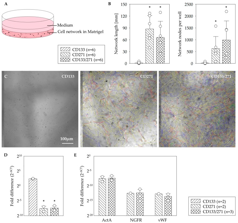
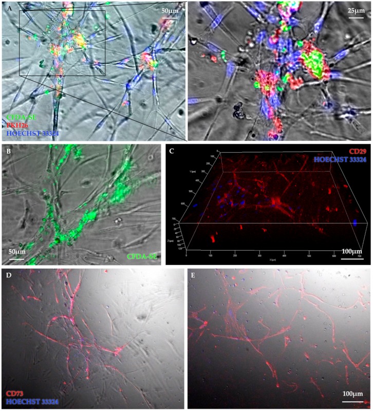
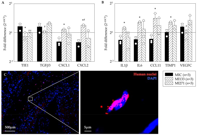
Methods and results: The effects of respective CD133+ and CD271+ mononuclear cells alone as well as in the co-culture model have been explored with focus on their angiogenic potential. The phenotypic analysis revealed a small percentage of isolated cells expressing both surface markers. Moreover, target stem cells isolated with our standardized immunomagnetic isolation procedure did not show any negative alterations following BM storage in regard to cell numbers and/or quality. In vitro network formation relied predominantly on CD271+ stem cells when compared with single CD133+ culture. Interestingly, CD133+ cells contributed in the tube formation, only if they were cultivated in combination with CD271+ cells. Additional to the in vitro examination, therapeutic effects of the primed stem cells were investigated 48 h post MI in a murine model. Hence, we have found a lower expression of transforming growth factor βeta 3 (TGFβ3) as well as an increase of the proangiogenic factors after CD133+ cell treatment in contrast to CD271+ cell treatment. On the other hand, the CD271+ cell therapy led to a lower expression of the inflammatory cytokines.

Conclusion: The interactions between CD271+ and CD133+ subpopulations the extent to which the combination may enhance cardiac regeneration has still not been investigated so far. We expect that the multiple characteristics and various regenerative effects of CD271+ cells alone as well as in combination with CD133+ will result in an improved therapeutic impact on ischemic heart disease.

Keywords: angiogenesis; bone marrow stem cells; cardiac regeneration; myocardial infarction.


Conflict of interest statement

The authors declare that they have no conflict of interest.

Figures

Figure 1
Figure 1
Quality of the immunomagnetic cell separation of CD271+ and CD133+ cells from fresh bone marrow (BM). Mean cell numbers of mononuclear cells (MNC), CD133+, and CD271+ cells achieved by magnet activated cell sorting (MACS) direct cell isolation (A). Linear dependence of CD133+ as well as CD271+ cell numbers on the initial amount of MNC (linear regression plots: RCD133 = 0.821; RCD271 = 0.689; B). The percentage of the CD133/CD271 double-positive cells determined within the different populations MNC, CD133+, as well as CD271+ cells using flow cytometry (C).
Figure 2
Figure 2
In vitro angiogenesis assay. Schematic illustration of the network formation assay within the Matrigel for testing of the angiogenesis potential (A). In contrast to CD133+ cells, a significant formation of network length and highest number of nodal points in the network of CD271+ cells as well as CD133+/CD271+ cell co-culture were observed (B). Representative phase contrast z-stack images of the mono- and co-culture assays (C). CD133+ cell culture in Matrigel led to a 10-fold higher level of ActA gene in comparison to CD133+/271+ cell co-culture (D). No differences were observed between CD271+ and CD133+/271+ cell culture assays for ActA, NGFR, and vWF gene expression (E). Mean ± SD; * p ≤ 0.015 vs. CD133, Mann–Whitney U test.
Figure 3
Figure 3
Representative images illustrate cellular interactions within the network structure. Colocalization of CFDA-SE labelled CD133+ cells (green) and PKH26-labelled CD271+ cells (red) at nodal points of the CD133+/CD271+ cell co-culture angiogenesis assay but with very rare or no connections of CD133+ cell culture were observed (A). Although CD133+ cell cultures built minimal networks and true CFDA-SE fluorescent signal originate inside the network structures (B). 3D-image of a vital mesenchymal cell network was taken 2 weeks after seeding (C). CD271+ cell (D) as well as CD133+/CD271+ cell co-culture (E) angiogenesis assays were stained with CD73-PE antibody (red) and counterstained for nuclei with Hoechst dye (blue).
Figure 4
Figure 4
Angiogenic benefit 48 h after myocardial infarction (MI). Data show a significant lower expression of fibrotic markers as well as significant improvement of pro angiogenic factors after CD133+ cell treatment compared to MIC as well as MI271 group (A). Lower expression of inflammatory cytokines after CD271+ stem cell treatment in contrast to significantly increase after CD133+ stem cell treatment as well as significantly improvement of the pro angiogenic factor VEGFC (vascular endothelial growth factor) after CD271+ cell treatment. (B). Mean ± SD normalized to SHAM operation; * p ≤ 0.05 vs. MIC; # p ≤ 0.05 vs. MI271 (t-test). Representative images of CD271+ cell derivatives illustrate the engrafted human stem cells 48 h post transplantation (C).

Similar articles

Cited by

References

    1. Donndorf P., Steinhoff G. CD133-Positive Cells for Cardiac Stem Cell Therapy: Current Status and Outlook. Adv. Exp. Med. Biol. 2013;777:215–227. doi: 10.1007/978-1-4614-5894-4_14. - DOI - PubMed
    1. Pittenger M.F., Mackay A.M., Beck S.C., Jaiswal R.K., Douglas R., Mosca J.D., Moorman M.A., Simonetti D.W., Craig S., Marshak D.R. Multilineage Potential of Adult Human Mesenchymal Stem Cells. Science. 1999;284:143–147. doi: 10.1126/science.284.5411.143. - DOI - PubMed
    1. Elnakish M.T., Kuppusamy P., Khan M. Stem Cell Transplantation as a Therapy for Cardiac Fibrosis. J. Pathol. 2013;229:347–354. doi: 10.1002/path.4111. - DOI - PubMed
    1. Jain M., Pfister O., Hajjar R.J., Liao R. Mesenchymal Stem Cells in the Infarcted Heart. Coron. Artery Dis. 2005;16:93–97. doi: 10.1097/00019501-200503000-00003. - DOI - PubMed
    1. Sánchez-Guijo F., Caballero-Velázquez T., López-Villar O., Redondo A., Parody R., Martinez C., Olavarria E., Andreu E., Prosper F., Diez-Campelo M., et al. Sequential Third-Party Mesenchymal Stromal Cell Therapy for Refractory Acute Graft-Versus-Host Disease. Biol. Blood Marrow Transplant. 2014;20:1580–1585. doi: 10.1016/j.bbmt.2014.06.015. - DOI - PubMed

MeSH terms