Review
. 2019 Dec 27;25(1):117.
doi: 10.3390/molecules25010117.

The Biosurfactant β-Aescin: A Review on the Physico-Chemical Properties and Its Interaction with Lipid Model Membranes and Langmuir Monolayers

Affiliations
Review

The Biosurfactant β-Aescin: A Review on the Physico-Chemical Properties and Its Interaction with Lipid Model Membranes and Langmuir Monolayers

Ramsia Geisler et al. Molecules. .

Abstract

This review discusses recent progress in physicochemical understanding of the action of the saponin β -aescin (also called β -escin), the biologically active component in the seeds of the horse chestnut tree Aesculus hippocastanum. β -Aescin is used in pharmacological and cosmetic applications showing strong surface activity. In this review, we outline the most important findings describing the behavior of β -aescin in solution (e.g., critical micelle concentration ( c m c ) and micelle shape) and special physicochemical properties of adsorbed β -aescin monolayers at the air-water and oil-water interface. Such monolayers were found to posses very special viscoelastic properties. The presentation of the experimental findings is complemented by discussing recent molecular dynamics simulations. These simulations do not only quantify the predominant interactions in adsorbed monolayers but also highlight the different behavior of neutral and ionized β -aescin molecules. The review concludes on the interaction of β -aescin with phospholipid model membranes in the form of bilayers and Langmuir monolayers. The interaction of β -aescin with lipid bilayers was found to strongly depend on its c m c . At concentrations below the c m c , membrane parameters are modified whereas above the c m c , complete solubilization of the bilayers occurs, depending on lipid phase state and concentration. In the presence of gel-phase phospholipids, discoidal bicelles form; these are tunable in size by composition. The phase behavior of β -aescin with lipid membranes can also be modified by addition of other molecules such as cholesterol or drug molecules. The lipid phase state also determines the penetration rate of β -aescin molecules into lipid monolayers. The strongest interaction was always found in the presence of gel-phase phospholipid molecules.

Keywords: Aesculus hippocastanum; Langmuir monolayers; air–water interface, MD simulation; critical micelle concentration cmc; lipid membrane; saponin; β-aescin; β-escin.


Conflict of interest statement

The authors declare no conflict of interest.

Figures

Figure 1
Figure 1
Number of publications related to the saponin β-aescin in the years 1967 to 2019. The data is obtained from a search in the ”The Web of Science Core Collection” database under the keyword (a)escin.
Figure 2
Figure 2
Molecular structure of β-aescin [29]. The amphiphilic structure of β-aescin consists of a hydrophobic, aglyconic part and a hydrophilic, glyconic part. The glyconic part contains glucuronic acid (GlcA), and glucose (Glc) (or xylose (Xyl)). However, in addition to the classical surfactant-like polarity, also within the aglyconic part polar groups are attached one-sided to the triterpene. This gives the aglyconic backbone a slightly polar and a slightly apolar side (Used abbreviations: Ang → angelic acid, Tig → tiglic acid).
Figure 3
Figure 3
TEM images of dry and stained β-aescin micelles. The concentrations of the solutions are (a) 2.5 mM and (b) 5.5 mM and were prepared at 6 C. Reproduced with permission from Dargel et al. [29], Colloids Interfaces; published by MDPI, 2019.
Figure 4
Figure 4
Proposed structure of saponin adsorption layers (AC) for a monodesmosidic saponin such as β-aescin at (A) water–air and (B,C) water–oil (hexadecane and tricaprylin) interfaces. Reproduced from Reference [44] with permission from The Royal Society of Chemistry.
Figure 5
Figure 5
The figure shows in (A) the gas permeability (Kfoam) as a function of pH of the solution for foams stabilized by either the horse chestnut tree extract (HC, blue squares) and the pure saponin β-aescin (red dots). In (B) the surface stress is plotted against the surface deformation for β-aescin adsorption layers formed from solutions with natural pH (pH = 3.1) and adjusted pH of 8.0. Reprinted from Colloids and Surfaces A, 534, Tcholakova et al. [40], Role of surface properties for the kinetics of bubble Ostwald ripening in saponin-stabilized foams, pp. 16–25, ©2017, with permission from Elsevier.
Figure 6
Figure 6
Schematic of the proposed orientation of densely packed β-aescin molecules at the water–air interface, as determined from NR experiments. Reprinted with permission from Penfold et al. [38], ©2018 American Chemical Society.
Figure 7
Figure 7
Proposed arrangement of β-aescin molecules of (A) neutral and (B) ionized molecules in a surface cluster adsorbed at a water-vacuum interface, as obtained from MD simulations. The numbers 1–4 indicate the main inter-molecular forces dominating the β-aescin self-assembly. These are: (1) long-range attraction due to the inhomogeneous charge distribution in the aglycone and short-range dispersion (London) van der Waals forces between the aglycone fragments; (2) intermediate-range dipole-dipole/H-bond interaction, (3) short-range classical H-bonds, and (4) water-screened electrostatic repulsion between the charged carboxyl groups in the ionized form of β-aescin molecules. Reprinted with permission from Tsibranska et al. [42], ©2017 American Chemical Society.
Figure 8
Figure 8
Summary of β-aescin-DMPC interactions depending on saponin concentration. The incorporation of β-aescin molecules into the DMPC bilayer increases the bilayer fluidity and β-aescin molecules phase-segregate into saponin-rich and saponin-poor domains in the membrane. With increasing β-aescin concentration, vesicles start to become unstable in solution and agglomerate due to the formation of larger domains. At β-aescin concentrations far above cmc of β-aescin (high conc. regime (III–IV)) the DMPC membrane is solubilized by the saponin molecules and discoidal bicelles form which are tunable in size by composition and temperature. Adapted with permission from Sreij et al. [46], ©2017 American Chemical Society.
Figure 9
Figure 9
Bilayer bending modulus κ as function of β-aescin content for 10 C and 40 C. Dotted lines are guides to the eye. Reproduced from Ref. [47] with permission from the PCCP Owner Societies.
Figure 10
Figure 10
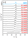
DSC of β-aescin containing samples. The red numbers denote β-aescin concentration in mol% with respect to the molar amount of used DMPC. An increasing β-aescin concentration enhances phase segregation and shows in the formation of a second peak at lower temperature. The experimental procedure is given elsewhere [21,48,55]. Adapted from Ref. [48] with permission of Elsevier.
Figure 11
Figure 11
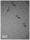
Cryo-TEM image of a sample with 4 mol% β-aescin prepared at 10 C. The arrows point on deformed SUVs (1), elongated SUVs (2), SUVs with edges (3) and stacks of bilayer disks (4). Reproduced from Ref. [48] with permission of Elsevier.
Figure 12
Figure 12
SANS (a) and SAXS (b) curves of β-aescin stabilized bicelles at 10 C for different aescin concentrations. The solid lines are fits obtained by using the IFT method developed by O. Glatter. (c) Normalized spatial correlation functions p(r) from SANS and (d) from SAXS from the respective panels (a) and (b). The legend indicates the β-aescin content in mol% with respect to the amount of DMPC. Reproduced with permission from Geisler et al. [49], ©2019 American Chemical Society.
Figure 13
Figure 13
TEM images of diverse samples. (A) β-aescin micelles in water. (BH): Pseudo-ternary and pseudo-binary systems prepared by the lipid film hydration method with aqueous solution. (B,C) DPPC:β-aescin:cholesterol mixture with a mass ratio of 1:3:1 (C) storage period of 2 months). (D) DPPC:β-aescin mixture with a mass ratio of 1:3. (E) cholesterol:β-aescin mixture with a mass ratio of 1:3. (F,G) TEM images of pseudo-ternary and pseudo-binary systems prepared by lipid film hydration method and hydration with Tris buffer (pH 7.4, 140 mM). (F) DPPC:β-aescin:cholesterol mixture with a mass ratio of 1:3: 1. G) DPPC:β-aescin mixture with a mass ratio of 1:3. (H) Cholesterol:β-aescin mixture with a mass ratio of 1:3. The scale bar equals to 200 nm. Reproduced from Reference [39] with permission of Planta Medica, © Georg Thieme Verlag KG.
Figure 14
Figure 14
Surface pressure-time adsorption kinetics for a DMPC monolayer after the start of buffer sub-phase exchange (50 mM phosphate buffer, pH 7.4) against β-aescin solution (same conditions) until reaching equilibrium pressure Πeq. The experiments were carried out at (a) 4 C and (b) 38 C for different initial pressures Πi 5, 15, and 25 mNm1. The arrows mark an intermediate cusp which is explained in the main text. Reproduced with permission from Sreij et al. [46], ©2017 American Chemical Society.
Figure 15
Figure 15
Results related to the surface pressure-time adsorption kinetics shown in Figure 14. The figure shows the insertion pressure (ΔΠ=ΠeqΠi) upon β-aescin incorporation into a DMPC monolayer as a function of initial pressure Πi obtained from Π(t) adsorption kinetics. The temperatures investigated are 4 C (triangles) and 38 C (dots). Filled symbols: LE phase; open symbols: LC phase. MIP (grey) denotes the maximum insertion pressure (MIP) at ΔΠ=0. Reproduced with permission from Sreij et al. [46], ©2017 American Chemical Society.

References

    1. Hostettmann K., Marston A. Saponins. Cambridge University Press; Cambridge, UK: New York, NY, USA: 1995. - DOI
    1. Güçlü-Üstündağ Ö., Mazza G. Saponins: Properties, applications and processing. Crit. Rev. Food Sci. Nutr. 2007;47:231–258. doi: 10.1080/10408390600698197. - DOI - PubMed
    1. Sparg S.G., Light M.E., van Staden J. Biological activities and distribution of plant saponins. J. Ethnopharmacol. 2004;94:219–243. doi: 10.1016/j.jep.2004.05.016. - DOI - PubMed
    1. Cheok C.Y., Salman H.A.K., Sulaiman R. Extraction and quantification of saponins: A review. Food Res. Int. 2014;59:16–40. doi: 10.1016/j.foodres.2014.01.057. - DOI
    1. Netala V.R., Ghosh S.B., Bobbu P., Anitha D., Tartte V. Triterpenoid saponins: A review on biosynthesis, applications and mechanism of their action. Int. Pharm. Pharm. Sci. 2015;7:24-8.

MeSH terms

LinkOut - more resources