Review
. 2019 Dec 31;26(1):4.
doi: 10.1186/s10020-019-0129-7.

Disorders of FZ-CRD; insights towards FZ-CRD folding and therapeutic landscape

Affiliations
Review

Disorders of FZ-CRD; insights towards FZ-CRD folding and therapeutic landscape

Reham M Milhem et al. Mol Med. .

Abstract

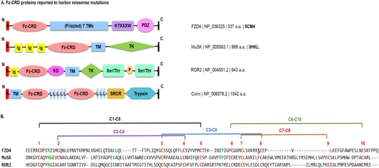
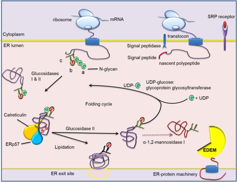
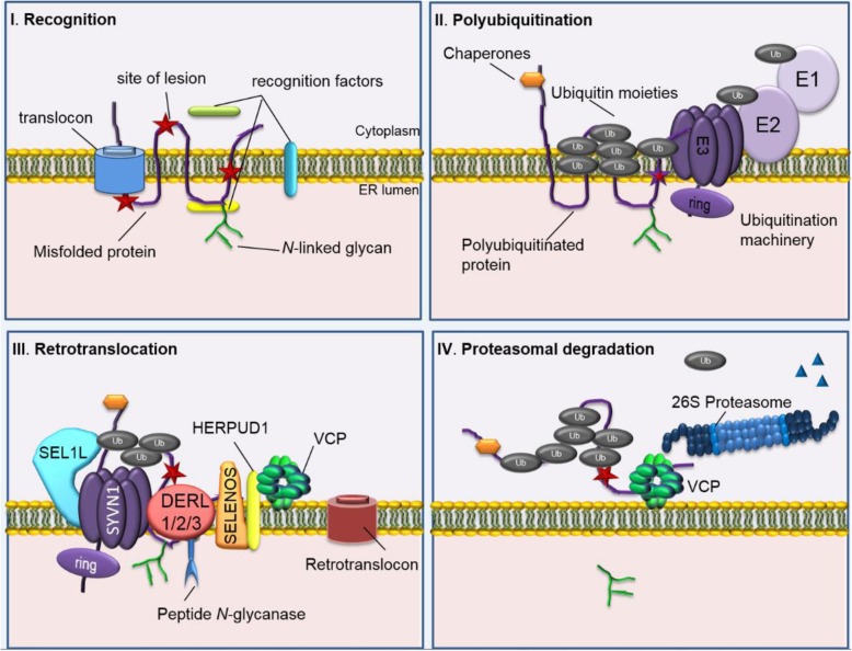
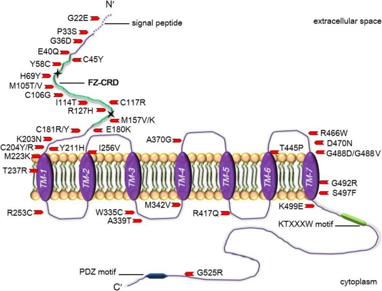
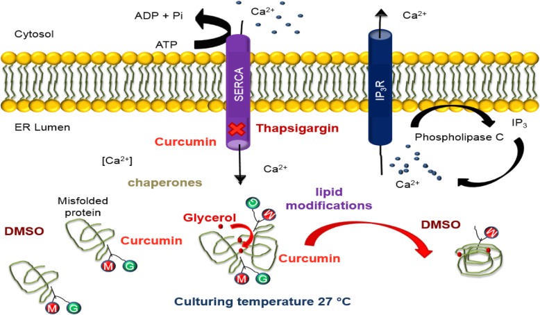
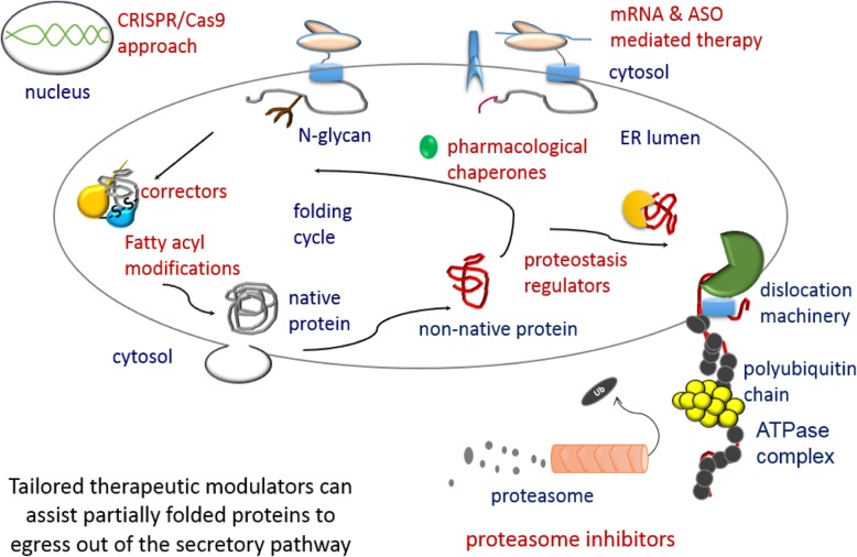
The ER is hub for protein folding. Proteins that harbor a Frizzled cysteine-rich domain (FZ-CRD) possess 10 conserved cysteine motifs held by a unique disulfide bridge pattern which attains a correct fold in the ER. Little is known about implications of disease-causing missense mutations within FZ-CRD families. Mutations in FZ-CRD of Frizzled class receptor 4 (FZD4) and Muscle, skeletal, receptor tyrosine kinase (MuSK) and Receptor tyrosine kinase-like orphan receptor 2 (ROR2) cause Familial Exudative Vitreoretinopathy (FEVR), Congenital Myasthenic Syndrome (CMS), and Robinow Syndrome (RS) respectively. We highlight reported pathogenic inherited missense mutations in FZ-CRD of FZD4, MuSK and ROR2 which misfold, and traffic abnormally in the ER, with ER-associated degradation (ERAD) as a common pathogenic mechanism for disease. Our review shows that all studied FZ-CRD mutants of RS, FEVR and CMS result in misfolded proteins and/or partially misfolded proteins with an ERAD fate, thus we coin them as "disorders of FZ-CRD". Abnormal trafficking was demonstrated in 17 of 29 mutants studied; 16 mutants were within and/or surrounding the FZ-CRD with two mutants distant from FZ-CRD. These ER-retained mutants were improperly N-glycosylated confirming ER-localization. FZD4 and MuSK mutants were tagged with polyubiquitin chains confirming targeting for proteasomal degradation. Investigating the cellular and molecular mechanisms of these mutations is important since misfolded protein and ER-targeted therapies are in development. The P344R-MuSK kinase mutant showed around 50% of its in-vitro autophosphorylation activity and P344R-MuSK increased two-fold on proteasome inhibition. M105T-FZD4, C204Y-FZD4, and P344R-MuSK mutants are thermosensitive and therefore, might benefit from extending the investigation to a larger number of chemical chaperones and/or proteasome inhibitors. Nonetheless, FZ-CRD ER-lipidation it less characterized in the literature and recent structural data sheds light on the importance of lipidation in protein glycosylation, proper folding, and ER trafficking. Current treatment strategies in-place for the conformational disease landscape is highlighted. From this review, we envision that disorders of FZ-CRD might be receptive to therapies that target FZ-CRD misfolding, regulation of fatty acids, and/or ER therapies; thus paving the way for a newly explored paradigm to treat different diseases with common defects.

Keywords: Cystic fibrosis conductance regulator protein; ERAD; protein misfolding; Familial exudative vitreoretinopathy; congenital myasthenic syndrome; Robinow syndrome; receptor tyrosine kinase-like orphan receptor 2; frizzled class receptor 4; muscle; Frizzled cysteine-rich domain; Frizzled receptors; Lipidation; Proteostasis; Receptor tyrosine kinase; conformational diseases; Skeletal; cis-unsaturated fatty acids.


Conflict of interest statement

The authors declare that they have no competing interests.

Figures

Fig. 1
Fig. 1
Reported FZD4 proteins with disease-causing missense proteins. a Protein domain structural models for FZ-CRD proteins. HUGO gene symbols proteins are shown next to the protein structure and the NCBI accession number is shown next to each protein model. Available PDB codes are in bold at the far right. [PDZ: PDZ binding motif, KTXXXW: lysine-threonine-X-X-X-tryptophan, TM: transmembrane, FZ-CRD: frizzled cysteine- rich domain, TK: tyrosine kinase domain, Ig: immunoglobulin domain, Ser/Thr: Serine-threonine/tyrosine-protein kinase, KD: kinase domain, Trypsin: trypsin-like protease domain, SPCR: scavenger receptor cysteine-rich domain, and L: low density lipoprotein receptor repeats. b Multiple sequence alignment of FZD4, MuSK and ROR2 FZ-CRDs. Conserved cysteines are shown in red color. FZ-CRD show homology with a conserved pattern of “CnCnCX8CX6CnCX3CX6,7CnCnC” (Pei and Grishin 2012) C: conserved cysteine; n: a variable number of residues, Xn: n residues, and Xn1, n2: n1 to n2 residues in α-helices forming a common Frizzled fold across four α-helices connected by disulfide bridges shown and labelled in red as: “C1–C5, C2–C4, C3–C8, C6–C10, and C7–C9” . For FZD4, one inserted region is shown as the number of inserted residues underlined in bold. Different residues exist between conserved C7 and C8. For FZDs the number is six and RTKs have seven residues
Fig. 2
Fig. 2
The glycoprotein folding cycle within the endoplasmic reticulum lumen. Protein glycosylation is a highly conserved process and plays crucial biological and physiological roles. Polypeptides translated on ribosomes from mRNA are escorted to an ER translocon via the signal recognition particle (SRP) and receptor. As the polypeptide enters the ER, an en bloc transfer N-glycans (Glc(3)Man(9)GlcNAc(2)) where glucose is represented as green circles and mannose as red, and N-acetylglucosamine (GlcNAc) is Y shaped green structure attached to the nascent polypeptide chain. FZD4 and MuSK have two N-glycosylation sites in their extracellular domains. α-glucosidase I and II (GI /GII) remove two of the three glucoses forming a monoglucosylated glycoprotein. This monoglucosylated protein is a signal for interacting with CNX and CRT, both lectins bound to protein disulfide isomerase family A member 3 (PDIA3). CRT is the soluble form of CNX and they form interchain disulfide bonds (S-S) with the bound glycoproteins. Removal of the last glucose by GII allows the glycoprotein to be released from the chaperones and leave the ER through ER exit sites to the golgi apparatus. Lipidation is a co or post-translational modification where lipid moieties are covalently attached to the polypeptide to increase hydrophobicity, conformation, and stability. Misfolded proteins trigger UDP-glucose-glucosyltransferase to re-add a single glucose on to the glycan and the cycle of protein folding is repeated. If the glycoprotein is permanently misfolded, the terminal mannose α1–2Man from the central arm of Man(9)GlcNAc(2), shown as a blue triangle, from the b branch of the oligosaccharide is removed by α-1,2-mannosidase I yielding a Man(8)GlcNAc(2) b-isomer. A second ER resident α-mannosidase I–like protein which lacks enzyme activity known as ER degradation-enhancing α-mannosidase I–like protein (EDEM), recognizes misfolded glycoproteins and targets them for ERAD machinery (Milhem 2015)
Fig. 3
Fig. 3
The four main steps for ERAD. I. Recognition occurs during protein synthesis. Here a misfolded region (red stars) are recognized by either cytoplasmic, ER luminal and/or transmembrane recognition factors depending on the site of lesion. II. Polyubiquitination starts when chaperones and co-chaperones direct the misfolded substrate to ubiquitination machinery. An ubiquitin activating enzyme (E1) transfers ubiquitin (Ub) (grey circles) to cysteine residue in an active site of an ubiquitin conjugating enzyme (E2) using ATP as energy. Ubiquitin ligase then transfers Ub to a lysine residue on the substrate protein. The latter process occurs on either the ER or cytoplasmic side of the membrane. III. Retrotranslocation ensues when the substrate protein is escorted to the dislocation machinery made up of a protein scaffold such as SEL1L adaptor subunit of ERAD E3 ubiquitin ligase (SEL1L), synoviolin 1 (SYVN1), cytochrome c oxidase assembly factor 7 (COA7) (not shown), derlin 1,2,3 (DERL1,2,3), selenoprotein S (SELENOS), homocysteine inducible ER protein with ubiquitin like domain 1 (HERPUD1), and valosin-containing protein (VCP). The substrate protein is removed either by passing through a retrotranslocon or by complete elimination of the protein. This is mainly done by the cytoplasmic ATPases associated with diverse cellular activities (AAA+ ATPase) p97 (commonly known as VCP), which interacts with Ub on the substrate and de-ubiquitinates the mutant protein and sends it off to the 26S proteasome. IV. Degradation is the final step where polyubiquitinated substrates are escorted to the 26S proteasome for degradation of faulty proteins. N-glycans are cleaved off by peptide N-glycanase associated with the ERAD machinery and Ub moieties are removed by de-ubuitinating enzymes found in the cytoplasm or in the proteasome cap to release small peptides shown as blue triangles (Milhem 2015)
Fig. 4
Fig. 4
Schematic representation showing 40 reported FZD4 missense mutations dispersed across the protein and are associated with pathogenic FEVR. FZD4 contains a signal sequence at the amino (N′) terminus from amino acids 1 and 36/37; a conserved FZ-CRD region highlighted in green of approximately 122 amino acids in the extracellular domain containing a motif of 10 spaced cysteines between amino acid positions 40 through 161; a seven-pass TMD region labelled TM1–7 within amino acid positions 210 through 514; and a cytoplasmic domain with a KTXXXW motif found at amino acid positions 499 through 504, and a PDZ motif located close to the C′ terminal at amino acid positions 535 through 537. The two potential N-glycosylation sites are indicated by black stars at amino acid positions 59 and 144. Smallwood et al. have shown that the binding of Norrin to the CRD domain of FZD4 extends to include residue C204 (Smallwood et al. 2007). Amino acid positions and domains can be accessed from https://www.uniprot.org/uniprot/Q9ULV1
Fig. 5
Fig. 5
The effects of targeting the intracellular environment of proteostasis. Glycerol has the ability to increase the hydration layer of the protein and the intramolecular hydrophobic bonding strength. This in turn allows the free movement of proteins in the crowded environment of the ER thereby preventing aggregation of proteins. Differing concentrations (0.1–1%) of DMSO in a cell may increase protein synthesis of the misfolded proteins or by possibly overwhelming the quality control system. Thapsigargin acts as an inhibitor of the Ca2+ ATP2A pump pump and increases cytosolic calcium, and in doing so results in an enhanced rescue of mutant proteins (Robben et al. 2006). Curcumin is a nontoxic natural constituent of turmeric spice and affects the Ca2+ ATP2A pump found on the ER plasma membrane. Curcumin inhibits the pumps ability to maintain a high ER Ca2+ level which disturbs the ability of ER molecular chaperones to target the misfolded protein for ERAD, hence, allows the mutant protein to exit the ER. Post-translational modifications of lipid modifications and glycosylation can be therapeutically targeted to support disulfide bond and glycoprotein formation to enhance the proteostasis network (Milhem 2015)
Fig. 6
Fig. 6
Summary: Therapeutically targeting the intracellular environment. The endoplasmic reticulum is a very important organelle for the proper folding of proteins that enter the secretory pathway. It contains stringent quality control checkpoints that monitor the folding of polypeptides and allow bone fide proteins to exit the ER and be expressed at their proper cellular localization. Research shows that proteins that are kinetically stable and thermostable in the ER, but do not conform to a proper conformation, can still progress to the secretory pathway and function similar to wild-type protein on the plasma membrane. Select therapeutic strategies are shown in red font. Pharmacological chaperones (correctors) can work at different levels of the folding cycle. Proteostasis regulators works with the ER quality control network and eliminate toxic non-native polypeptides. Fatty acyl modifications assist in proper cysteine bond formation and compact polypeptide folding. Abbreviations; ASO: antisense oligonucleotides, CRISPR/Cas9: clustered regulatory interspaced short palindromic repeats)/cas9 systems

Similar articles

Cited by

References

    1. Afzal AR, Rajab A, Fenske CD, Oldridge M, Elanko N, Ternes-Pereira E, et al. Recessive Robinow syndrome, allelic to dominant brachydactyly type B, is caused by mutation of ROR2. Nat Genet. 2000;25(4):419–422. doi: 10.1038/78107. - DOI - PubMed
    1. Ahner A, Brodsky JL. Checkpoints in ER-associated degradation: excuse me, which way to the proteasome? Trends Cell Biol. 2004;14(9):474–478. doi: 10.1016/j.tcb.2004.07.013. - DOI - PubMed
    1. Ali BR, Ben-Rebeh I, John A, Akawi NA, Milhem RM, Al-Shehhi NA, et al. Endoplasmic reticulum quality control is involved in the mechanism of endoglin-mediated hereditary haemorrhagic telangiectasia. PLoS One. 2011;6(10):e26206. doi: 10.1371/journal.pone.0026206. - DOI - PMC - PubMed
    1. Ali BR, Jeffery S, Patel N, Tinworth LE, Meguid N, Patton MA, et al. Novel Robinow syndrome causing mutations in the proximal region of the frizzled-like domain of ROR2 are retained in the endoplasmic reticulum. Hum Genet. 2007;122(3–4):389–395. doi: 10.1007/s00439-007-0409-0. - DOI - PubMed
    1. Anastas JN, Moon RT. WNT signalling pathways as therapeutic targets in cancer. Nat Rev Cancer. 2013;13(1):11–26. doi: 10.1038/nrc3419. - DOI - PubMed

Publication types

LinkOut - more resources