. 2020 Apr 15;87(8):745-755.
doi: 10.1016/j.biopsych.2019.10.025. Epub 2019 Nov 6.

Deletion of Glycogen Synthase Kinase-3β in D2 Receptor-Positive Neurons Ameliorates Cognitive Impairment via NMDA Receptor-Dependent Synaptic Plasticity

Affiliations

Deletion of Glycogen Synthase Kinase-3β in D2 Receptor-Positive Neurons Ameliorates Cognitive Impairment via NMDA Receptor-Dependent Synaptic Plasticity

Yan-Chun Li et al. Biol Psychiatry. .

Abstract

Background: Cortical dopaminergic systems are critically involved in prefrontal cortex (PFC) functions, especially in working memory and neurodevelopmental disorders such as schizophrenia. GSK-3β (glycogen synthase kinase-3β) is highly associated with cAMP (cyclic adenosine monophosphate)-independent dopamine D2 receptor (D2R)-mediated signaling to affect dopamine-dependent behaviors. However, the mechanisms underlying the GSK-3β modulation of cognitive function via D2Rs remains unclear.

Methods: This study explored how conditional cell-type-specific ablation of GSK-3β in D2R+ neurons (D2R-GSK-3β-/-) in the brain affects synaptic function in the medial PFC (mPFC). Both male and female (postnatal days 60-90) mice, including 140 D2R, 24 D1R, and 38 DISC1 mice, were used.

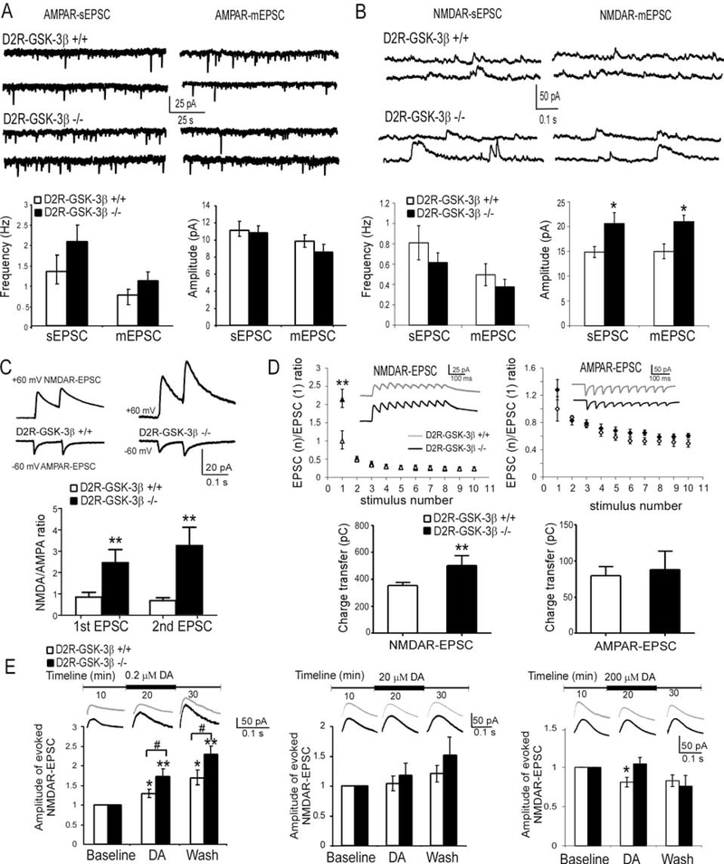
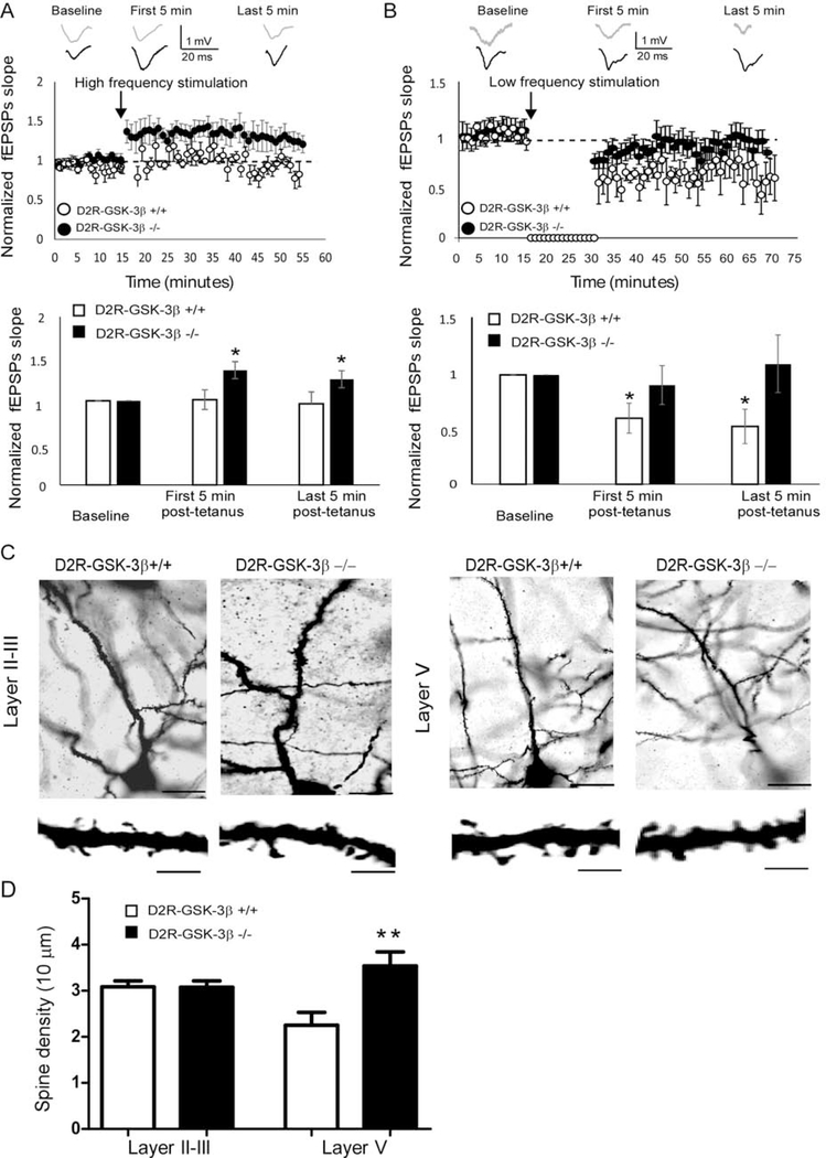
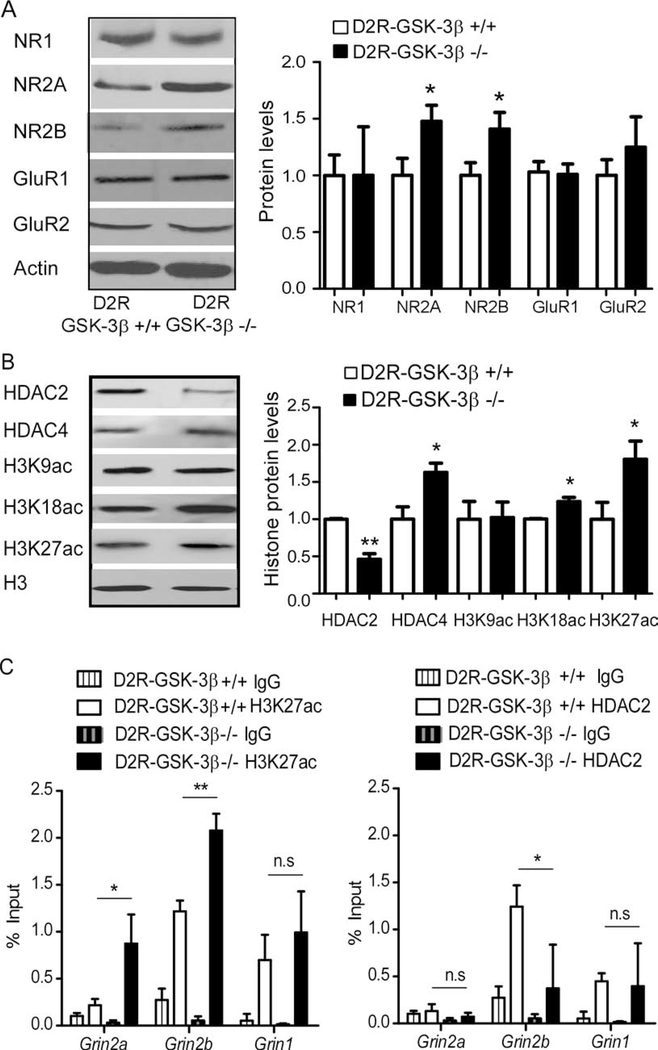
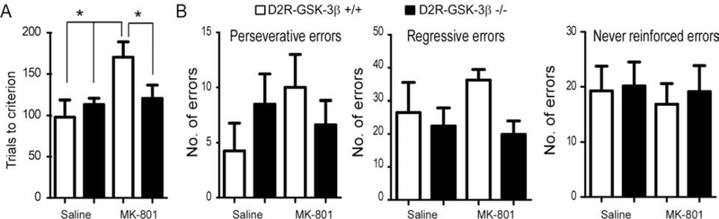
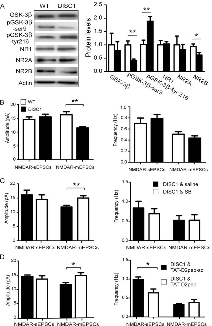
Results: This study found that NMDA receptor (NMDAR) function was significantly increased in layer V pyramidal neurons in mPFC of D2R-GSK-3β-/- mice, along with increased dopamine modulation of NMDAR-mediated current. Consistently, NR2A and NR2B protein levels were elevated in mPFC of D2R-GSK-3β-/- mice. This change was accompanied by a significant increase in enrichment of activator histone mark H3K27ac at the promoters of both Grin2a and Grin2b genes. In addition, altered short- and long-term synaptic plasticity, along with an increased spine density in layer V pyramidal neurons, were detected in D2R-GSK-3β-/- mice. Indeed, D2R-GSK-3β-/- mice also exhibited a resistance of working memory impairment induced by injection of NMDAR antagonist MK-801. Notably, either inhibiting GSK-3β or disrupting the D2R-DISC1 complex was able to reverse the mutant DISC1-induced decrease of NMDAR-mediated currents in the mPFC.

Conclusions: This study demonstrates that GSK-3β modulates cognition via D2R-DISC1 interaction and epigenetic regulation of NMDAR expression and function.

Keywords: Cognition; Dopamine D(2) receptors; Epigenetic; GSK-3β; Histone modification; NMDA receptors; Prefrontal cortex.


Conflict of interest statement

Conflict of Interests

The authors report no biomedical financial interests or potential conflicts of interest.

Figures

Figure 1:
Figure 1:
Deletion of GSK-3β in D2R+ (D2R-GSK-3β−/−) neurons in the brain enhanced NMDAR function and DA sensitivity in layer V pyramidal neurons of mouse mPFC. A, sample traces of AMPAR-sEPSCs and AMPAR-mEPSCs recorded from layer V pyramidal neurons in the PFC (upper panel). D2R-GSK-3β−/− mice did not show significant changes in AMPAR-EPSCs compared with their littermate controls (n = 25, p > 0.05 for both; lower panel). B, sample traces of NMDAR-sEPSCs and NMDAR-mEPSCs recorded from layer V pyramidal neurons in the PFC (upper panel). Summary histograms in the lower panel exhibited significant increases in amplitude (n = 20, *p < 0.05 for both sEPSCs and mEPSCs) but not the frequency of either NMDAR-sEPSCs or NMDAR-mEPSCs (n = 20, p > 0.05) in D2R-GSK-3β−/− mice. C, sample traces of evoked AMPAR-EPSC (−60 mV) and NMDAR-EPSC (+60 mV) recorded from the same neuron in the mPFC, and the histogram showed that deletion of GSK-3β in D2R+ neurons significantly increases both 1st and 2nd NMDA/AMPA ratios (vs. D2R-GSK-3β+/+, n = 8, **p < 0.01 for both 1st and 2nd EPSC). D, upper: NMDAR-EPSC or AMPAR-EPSC trains in D2R-GSK-3β−/− mice and their littermate controls. The EPSC currents elicited in the train were normalized to the amplitude of the first EPSC and were plotted against the stimulus numbers. The amplitude of the 1st NMDAR-EPSC but not AMPAR-EPSC was dramatically increased in D2R-GSK-3β−/− mice compared with their littermate controls (n = 10 for each group, ** P < 0.01 for NMDAR-EPSC and p > 0.05 for AMPAR-EPSC), but paired-pulse ratios (PPR) of neither NMDAR-EPSCs nor AMPAR-EPSCs elicited by repetitive pulses were changed (i.e., EPSC2–10, p > 0.05 for all). Lower: the charger transfer of NMDAR-EPSCs but not AMPAR-EPSC were significantly increased in D2R-GSK-3β−/− mice (n = 10 for each group, **p < 0.01 for NMDAR and p > 0.05 for AMPAR). E, Top panel: samples traces of NMDAR-EPSCs. Lower panels: left: at low dose of 0.2 μM, the enhancing effects of DA on NMDAR-EPSCs were potentiated during 10 min DA application and washing period in D2R-GSK-3β−/− mice (n = 8, *p < 0.05 for both DA wash-in and wash-out in D2R-GSK-3β+/+ mice; n = 8, **p < 0.01 in D2R-GSK-3β−/− mice; # p < 0.05 for both DA wash-in and wash-out in D2R-GSK-3β+/+ mice vs. D2R-GSK-3β−/− mice). Middle: at a higher dose of 20 μM, DA didn’t show any significant effects on NMDAR-EPSCs in both D2R-GSK-3β+/+ mice and D2R-GSK-3β−/− mice (n = 8 for both groups, p > 0.05). Right: at a high dose of 200 μM, the suppressive effect of DA found in D2R-GSK-3β+/+ mice was blunted in D2R-GSK-3β−/− mice (n = 8, *p < 0.05 for DA application in D2R-GSK-3β+/+ mice; n = 8, p > 0.05 for DA application in D2R-GSK-3β−/− mice; p > 0.05 for both DA wash-in and wash-out in D2R-GSK-3β+/+ mice vs. D2R-GSK-3β−/− mice).
Figure 2:
Figure 2:
Neuronal plasticity and dendritic spine were altered in D2R-GSK-3β−/− mice. A, LTP induction. Upper: representative fEPSP traces were recorded from mPFC layer V neurons of D2R-GSK-3β+/+ mice and D2R-GSK-3β−/− mice and graphical representation showing normalized fEPSP slope during baseline recording and following high-frequency (six trains of 100 pulse at 100 Hz) stimulation. Lower: summary histogram showed that the fEPSP slopes in both first 5 min and last 5 min after high-frequency stimulation were significantly increased in D2R-GSK-3β−/− mice, but not in D2R-GSK-3β+/+ mice, suggesting an increased LTP induction in D2R-GSK-3β−/− mice (n = 10, D2R-GSK-3β+/+: p > 0.05 for both the first and last 5 min post-tetanus; n = 10, D2R-GSK-3β−/−: * p < 0.05, both the first and last 5 min post-tetanus). B, LTD induction. Upper: representative fEPSP traces were recorded from mPFC layer V pyramidal neurons of D2R-GSK-3β+/+ mice and D2R-GSK-3β−/− mice and graphical representation showing normalized fEPSP slope during baseline recording and following low-frequency stimulation (900 pulses at 1Hz). Lower: summary histogram showed that the fEPSP slopes in both the first 5 min and last 5 min of low-frequency stimulation were decreased in wild-type D2R-GSK-3β+/+ mice, but the no LTD was induced in D2R-GSK-3β−/− mice, suggesting a terminated LTD in D2R-GSK-3β−/− mice (n = 8, D2R-GSK-3β+/+: *p < 0.05 for both the first and last 5 min post-tetanus; n = 8, D2R-GSK-3β−/−: p > 0.05, both the first and last 5 min post-tetanus). C, upper: Golgi–Cox-stained individual layers II-III and layer V pyramidal neurons in the mPFC from D2R-GSK-3β+/+ and D2R-GSK-3β−/− mice. High-magnification images of apical dendritic spines were shown in the lower panels. Scale bars = 50 μm for the upper panel and 10 μm for the lower panel. D, summary histogram showed that spine density in layer II-III and layer V pyramidal neurons of D2R-GSK-3β+/+ and D2R-GSK-3β−/− mice (n = 10 from 4 mice for each group, p > 0.05 for layer II-III and *p < 0.05 for layer V).
Figure 3:
Figure 3:
Histone modification is involved in the increase of NMDAR protein expression in mPFC of D2R-GSK3−/− mice. A, representative Western blots and summary histograms showed that total protein levels of NMDAR and AMPAR subunits from the mPFC of D2R-GSK-3β+/+ and D2R-GSK-3β−/− mice. Both NR2A and NR2B were significantly increased in D2R-GSK-3β−/− mice (n = 8 for each group, *p < 0.05 for both), but NR1, GluR1, and GluR2 were unchanged (n = 8 for each group, p > 0.05 for all). B, representative Western blots and summary histograms showed that total protein levels of HDAC2 & 4, as well as three acetylation sites of H3K from mPFC tissue of D2R-GSK-3β+/+ mice and D2R-GSK-3β−/− mice. HDAC2 was decreased while HDAC4 was increased in D2R-GSK-3β−/− mice (vs. D2R-GSK-3β−/− mice, n = 6 for each group, *p < 0.05 for both). Both H3K18ac and H3K27ac, but not H3K9ac, were increased in D2R-GSK-3β−/− mice (vs. D2R-GSK-3β−/− mice, n = 6 for each group, *p < 0.05). C, chromatin was pooled from the PFC of 2 animals from D2R-GSK-3β+/+ or D2R-GSK-3β−/− mice and immunoprecipitated using antibodies against H3K27ac (left) and HDAC2 (right). qPCR was performed using a primer set specific to the promoter region of Grin1, Grin2a, and Grin2b. The signal of the amplified DNA was normalized to input. H3K27ac enrichment at either Grin2a or Grin2b but not Grin1 were significantly enhanced in D2R-GSK-3β−/− (vs. D2R-GSK-3β+/+, n = 6 mice yielding 3 data points for each group, *p < 0.05, **p < 0.01). On the contrary, HDAC2 enrichment at Grin2b, but not Grin1 and Grin2a, was significantly reduced in D2R-GSK-3β−/− mice (vs. D2R-GSK-3β+/+, n = 6 mice yield 3 data points for each group, *p < 0.05).
Figure 4:
Figure 4:
D2R-GSK-3β−/− mice resisted MK-801-induced working memory deficits. A, two-way ANOVA analysis of the number of trials for D2R-GSK-3β+/+ mice and D2R-GSK-3β−/− mice to reach criteria revealed a significant difference in interaction effect (n = 8 for each group, F = 6.28, *p < 0.05). The further simple effect test analysis showed that the number of trials significantly increased after injection of MK-801 in D2R-GSK-3β+/+ mice compared with either injection of saline in D2R-GSK-3β+/+ mice or D2R-GSK-3β−/− mice (*p < 0.05 for both). However, injection of MK-801 in D2R-GSK-3β−/− mice exhibited less number of trials compared with that in D2R-GSK-3β+/+ mice (n = 8 for each group, *p < 0.05). B, there were no significant differences in three types of error analyzed among D2R-GSK-3β+/+ saline, D2R-GSK-3β+/+ MK-801, D2R-GSK-3β−/− saline and D2R-GSK-3β−/− MK-801 (Perseverative errors: interaction F = 1.78, p > 0.05; treatment F = 0.46 p > 0.05; genotypes F = 0.02, p > 0.05. Regressive errors: interaction F = 2.66, p > 0.05; treatment F = 0.12, p > 0.05; genotypes F = 2.89, p >0.05. Never reinforced errors: interaction F = 0.02, p > 0.05; treatment F = 0.14, p > 0.05; genotypes F = 0.12, p > 0.05).
Figure 5:
Figure 5:
Inhibiting GSK-3β or interrupting the interaction of D2R and DISC1 restored NMDAR function in mutant hDISC1 mice. A, representative Western blots and summary histograms showed that total protein levels of GSK-3β, pGSK-3β, and NMDAR subunits from mPFC tissue of mutant hDISC1 mice and their littermate controls. pGSK-3β at serine 9 (pGSK-3β-Ser9) was decreased while pGSK3β tyrosine 216 (pGSK-3β-Tyr216) was increased in mutant hDISC1 overexpressing mice compared with control mice (n = 6 for each group, **p < 0.01 for both). NR2B was reduced in mutant hDISC1 mice compared with control mice (n = 6 for each group, *p < 0.05). There were no significant differences in GSK-3β, NR1, and NR2A detected between mutant hDISC1 mice and control mice (n = 6 for each group, p > 0.05 for all). B, NMDAR-sEPSCs, and NMDAR-mEPSCs were recorded from layer V pyramidal neurons in the mPFC of both mutant hDISC1 mice and their littermate controls. The amplitude but not the frequency of NMDAR-mEPSCs was significantly decreased in mutant hDISC1 mice compared with control mice (n = 6 for each group, *p < 0.05). C, treatment with selective GSK-3β inhibitor SB216763 (2 mg/kg/day, i.p., once a day for 5 days) during juvenile period prevented a decrease in NMDAR-mEPSC amplitude compared with saline injection in mutant hDISC1 mice (n = 12 for each group, *p < 0.05). D, similarly, treatment with TAT-D2pep (10 μM, i.p., once a day for 5 days) during the juvenile period also prevents a decrease in NMDAR-mEPSC amplitude (but not frequency) compared with scrambled control peptide (TAT-D2pep-sc) treated mutant hDISC1 mice (n = 10 for each group, *p < 0.05). However, treatment with TAT-D2pep also caused a significant decrease in the frequency of NMDAR-sEPSC compared with scrambled control peptide (TAT-D2pep-sc) treated mutant hDISC1 mice (n = 10 for each group, *p < 0.05).

Comment in

References

    1. Seong HJ, Carter AG (2012): D1 receptor modulation of action potential firing in a subpopulation of layer 5 pyramidal neurons in the prefrontal cortex. The Journal of neuroscience : the official journal of the Society for Neuroscience. 32:10516–10521. - PMC - PubMed
    1. Gee S, Ellwood I, Patel T, Luongo F, Deisseroth K, Sohal VS (2012): Synaptic activity unmasks dopamine D2 receptor modulation of a specific class of layer V pyramidal neurons in prefrontal cortex. The Journal of neuroscience : the official journal of the Society for Neuroscience. 32:4959–4971. - PMC - PubMed
    1. Seamans JK, Yang CR (2004): The principal features and mechanisms of dopamine modulation in the prefrontal cortex. Prog Neurobiol. 74:1–58. - PubMed
    1. Missale C, Nash SR, Robinson SW, Jaber M, Caron MG (1998): Dopamine receptors: from structure to function. Physiological reviews. 78:189–225. - PubMed
    1. Greengard P (2001): The neurobiology of slow synaptic transmission. Science. 294:1024–1030. - PubMed

Publication types

Substances