. 2020 Jan 21;118(2):366-375.
doi: 10.1016/j.bpj.2019.12.011. Epub 2019 Dec 18.

Rapid Simulation of Unprocessed DEER Decay Data for Protein Fold Prediction

Affiliations

Rapid Simulation of Unprocessed DEER Decay Data for Protein Fold Prediction

Diego Del Alamo et al. Biophys J. .

Abstract

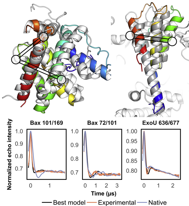
Despite advances in sampling and scoring strategies, Monte Carlo modeling methods still struggle to accurately predict de novo the structures of large proteins, membrane proteins, or proteins of complex topologies. Previous approaches have addressed these shortcomings by leveraging sparse distance data gathered using site-directed spin labeling and electron paramagnetic resonance spectroscopy to improve protein structure prediction and refinement outcomes. However, existing computational implementations entail compromises between coarse-grained models of the spin label that lower the resolution and explicit models that lead to resource-intense simulations. These methods are further limited by their reliance on distance distributions, which are calculated from a primary refocused echo decay signal and contain uncertainties that may require manual refinement. Here, we addressed these challenges by developing RosettaDEER, a scoring method within the Rosetta software suite capable of simulating double electron-electron resonance spectroscopy decay traces and distance distributions between spin labels fast enough to fold proteins de novo. We demonstrate that the accuracy of resulting distance distributions match or exceed those generated by more computationally intensive methods. Moreover, decay traces generated from these distributions recapitulate intermolecular background coupling parameters even when the time window of data collection is truncated. As a result, RosettaDEER can discriminate between poorly folded and native-like models by using decay traces that cannot be accurately converted into distance distributions using regularized fitting approaches. Finally, using two challenging test cases, we demonstrate that RosettaDEER leverages these experimental data for protein fold prediction more effectively than previous methods. These benchmarking results confirm that RosettaDEER can effectively leverage sparse experimental data for a wide array of modeling applications built into the Rosetta software suite.


Figures

Figure 1
Figure 1
Simulations of distance distributions between nitroxide probes using RosettaDEER. (A) An example of an experimentally observed distance distribution in apo Mhp1 51/278, shown in black. Distance distributions were simulated using RosettaDEER, MMM, and MDDS from the occluded Mhp1 structure (PDB: 2JLN). The average distance between Cβ atoms and the average distance calculated using the CONE model shown in light gray and dark gray, respectively. (B) The estimated average time required to simulate distance distributions (the lower limit of quantitation exceeded the Cβ-Cβ distance compute time). (C) Coarse-grained rotameric ensemble representation of the methanethiosulfonate spin label. Centers of mass, shown in purple, are used for clash evaluation, whereas electron coordinates, shown in gray, serve as measurement coordinates. (D) Distance distributions between residues are simulated by superimposing coordinates, evaluating clashes and measuring all resulting pairwise distances.
Figure 2
Figure 2
RosettaDEER simulations of distance distributions and decay traces. The forward approach taken by RosettaDEER contrasts with the preprocessing required by the CONE model. (A) A flowchart illustrating how both the CONE model and RosettaDEER use experimental DEER data to model proteins (example shown is T4 lysozyme residues 93 and 123). (B) Incorporation of DEER experimental restraints into Rosetta structure prediction pipeline. (C) Recovery of experimental background coupling and modulation depth parameter values.
Figure 3
Figure 3
Evaluation of models using DEER decay traces. Models with Cα RMSD100SSE ranging from 0.5 Å to 20.0–30.0 Å were scored using both the Rosetta energy function and RosettaDEER.
Figure 4
Figure 4
Structure prediction of Bax and ExoU using DEER decay data. (A) De novo protein folding of native-like models using DEER decay restraints with RosettaDEER, Cβ-Cβ distance restraints with the CONE model, or no restraints. Inset: spread of all models generated using these three methods. (B) Accuracy of de novo folded models (gray dots) and clusters (black circles) as a function of combined DEER and Rosetta Z-score.
Figure 5
Figure 5
Predicted models of Bax and ExoU at 3.2 and 2.1 Å Cα RMSD100SSE. (Top) Models were obtained from 10,000 de novo folded models, the best-scoring of which were refined into 1200 additional models. Native models shown in white. (Bottom) Example DEER traces in which the best model outperformed the native. Corresponding residues indicated as circles in (A) and (B).

References

    1. Steven A.C., Baumeister W. The future is hybrid. J. Struct. Biol. 2008;163:186–195. - PubMed
    1. Xia Y., Fischer A.W., Meiler J. Integrated structural biology for α-Helical membrane protein structure determination. Structure. 2018;26:657–666.e2. - PMC - PubMed
    1. Jeschke G. The contribution of modern EPR to structural biology. Emerg. Top. Life Sci. 2018;2:9–18. - PMC - PubMed
    1. Sahu I.D., Lorigan G.A. Site-directed spin labeling EPR for studying membrane proteins. BioMed Res. Int. 2018;2018:3248289. - PMC - PubMed
    1. Alexander N., Bortolus M., Meiler J. De novo high-resolution protein structure determination from sparse spin-labeling EPR data. Structure. 2008;16:181–195. - PMC - PubMed

Publication types