. 2019 Dec 31;9(1):20392.
doi: 10.1038/s41598-019-56932-8.

Intraoperative assessment of skull base tumors using stimulated Raman scattering microscopy

Affiliations

Intraoperative assessment of skull base tumors using stimulated Raman scattering microscopy

Kseniya S Shin et al. Sci Rep. .

Abstract

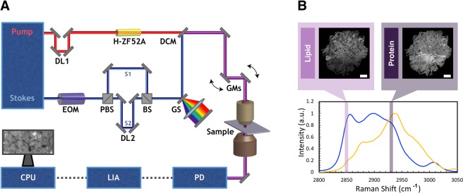
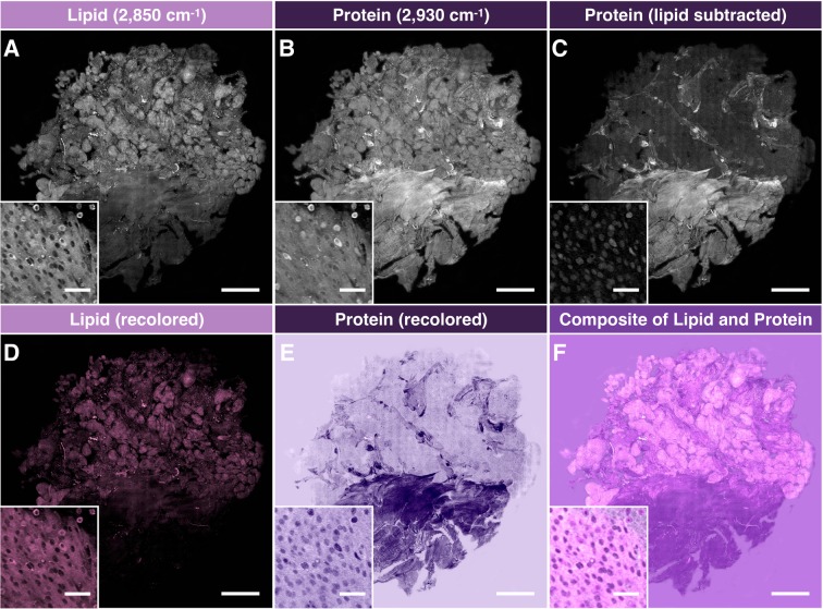
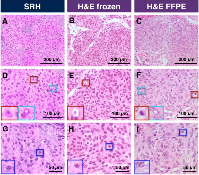
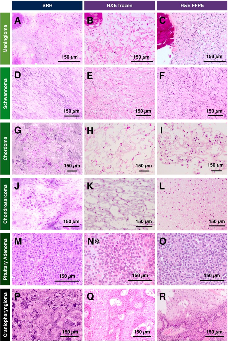
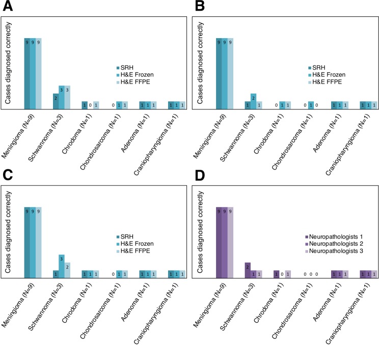
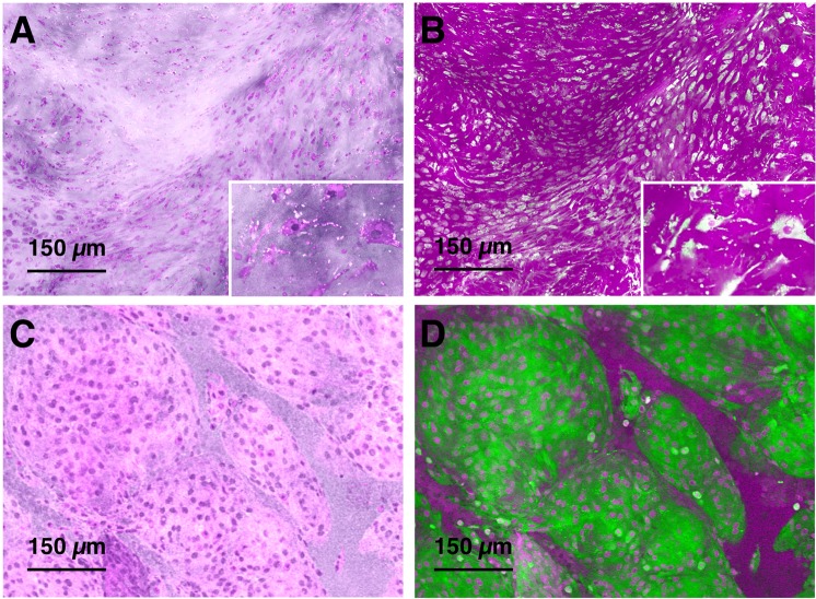
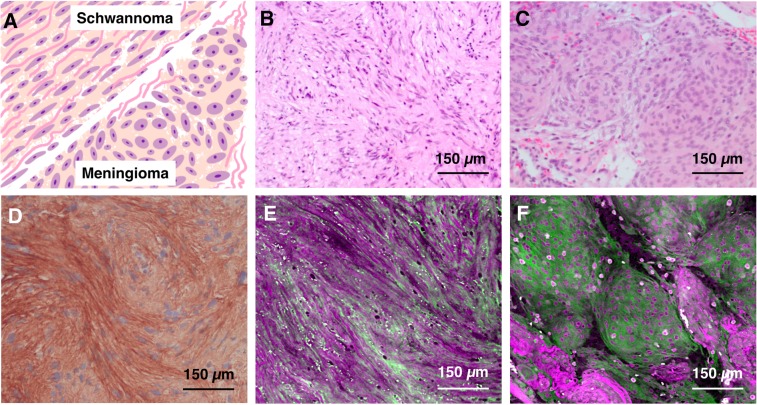
Intraoperative consultations, used to guide tumor resection, can present histopathological findings that are challenging to interpret due to artefacts from tissue cryosectioning and conventional staining. Stimulated Raman histology (SRH), a label-free imaging technique for unprocessed biospecimens, has demonstrated promise in a limited subset of tumors. Here, we target unexplored skull base tumors using a fast simultaneous two-channel stimulated Raman scattering (SRS) imaging technique and a new pseudo-hematoxylin and eosin (H&E) recoloring methodology. To quantitatively evaluate the efficacy of our approach, we use modularized assessment of diagnostic accuracy beyond cancer/non-cancer determination and neuropathologist confidence for SRH images contrasted to H&E-stained frozen and formalin-fixed paraffin-embedded (FFPE) tissue sections. Our results reveal that SRH is effective for establishing a diagnosis using fresh tissue in most cases with 87% accuracy relative to H&E-stained FFPE sections. Further analysis of discrepant case interpretation suggests that pseudo-H&E recoloring underutilizes the rich chemical information offered by SRS imaging, and an improved diagnosis can be achieved if full SRS information is used. In summary, our findings show that pseudo-H&E recolored SRS images in combination with lipid and protein chemical information can maximize the use of SRS during intraoperative pathologic consultation with implications for tissue preservation and augmented diagnostic utility.


Conflict of interest statement

The authors declare no competing interests.

Figures

Figure 1
Figure 1
Experimental set up for SRS microscope and controls for lipid and protein channels. (A) The Stokes laser is modulated by an electro-optical modulator (EOM) at 20 MHz. Tunable laser is spectrally dispersed through glass rods (tunable) and Stokes laser is dispersed through a grating stretcher (GS). They are the spatially and temporally overlapped at a dichroic mirror (DCM) and directed onto a pair of galvanomirrors (GM). The beams are then sent through a laser scanning microscope with a 25x water immersion objective. The pump beam after the condenser is detected by a photodiode (PD). The signal is processed through lock-in amplifier (LIA). The images are collected using ScanImage on computer processing unit (CPU). (B) SRS spectra for oleic acid and bovine serum albumin (BSA) as controls for lipids and proteins, respectively.
Figure 2
Figure 2
Image processing of stitched SRS imaging data (Meningioma, WHO grade I). (A) Stitched field-normalized data for lipid channel. (B) Stitched field-normalized data for protein channel. (C) Lipid data subtracted from protein, utilizing lipid and protein images in (A,B). (D) Recoloring result of lipid data. (E) Recoloring result of protein data. (F) Composite image of (D,E). Whole tissue scale bar: 1 mm. Inset scale bar: 50 μm.
Figure 3
Figure 3
Comparison of cellular features available with SRH versus conventional H&E stained slides in representative case of Meningioma WHO grade I. (A–C) Comparison of architecture features. (D–F) Increased magnification highlighting macrophages (red box) verified by immunohistochemistry and intranuclear inclusions (cyan box). (G–I) Nuclear features with easily identifiable nucleolus (blue box).
Figure 4
Figure 4
Comparison of SRH with conventional histological preparations of skull base tumors. (A–C) Meningioma, WHO grade I. (D–F) Schwannoma, WHO grade I. (G–I) Chordoma. (J–L) Chondrosarcoma, grade 2. (M,N*,O) Sparsely granulated somatotroph adenoma. (P–R) Papillary craniopharyngioma (BRAF-mutant). *The case warranted cytological preparations only during intraoperative consultation.
Figure 5
Figure 5
Depiction of correctly diagnosed cases for each neuropathologist and modality. (A–C) Neuropathologist 1, 2, and 3, respectively. (D) SRH only comparison for all neuropathologists.
Figure 6
Figure 6
Chondrosarcoma versus meningioma. (A) Pseudo-H&E recolored SRH of chondrosarcoma case. (B) Lipid (green) and protein (magenta) SRS of chondrosarcoma case. (C) Pseudo-H&E recolored SRH of meningioma case. (D) Lipid (green) and protein (magenta) SRS of meningioma case.
Figure 7
Figure 7
SRS images improve the identification of collagen. (A) Depiction of architectural and morphological differences between meningioma and schwannoma with particular focus on collagen fibers. (B) H&E of schwannoma. (C) H&E of meningioma. Protein signal is shown in magenta while lipid signal is shown as green. (D) Sample of collagen type IV antibody stained schwannoma case. (E) SRS based image of schwannoma. (F) SRS based image of meningioma.

References

    1. Powell SZ. Intraoperative Consultation, Cytologic Preparations, and Frozen Section in the Central Nervous System. Arch Pathol Lab Med. 2005;129:18. - PubMed
    1. Chatterjee S. Artefacts in histopathology. J. Oral Maxillofac. Pathol.: JOMFP. 2014;18:S111–S116. doi: 10.4103/0973-029X.141346. - DOI - PMC - PubMed
    1. Louis, D., Ohgaki, H., Wiestler, O. & Cavenee, W. (eds) WHO Classification of Tumours of the Central Nervous System, Fourth Edition fourth edn, (World Health Organization and International Agency for Research on Cancer, 2007).
    1. Mazzoni A, Krengli M. Historical development of the treatment of skull base tumours. Reports Pract. Oncol. Radiother. 2016;21:319–324. doi: 10.1016/j.rpor.2014.12.001. - DOI - PMC - PubMed
    1. Jaju H. Unfavourable results in skull base surgery. Indian J. Plast. Surg.: Off. Publ. Assoc. Plast. Surg. India. 2013;46:239–246. doi: 10.4103/0970-0358.118599. - DOI - PMC - PubMed

MeSH terms