Review
. 2020 Mar;122(6):735-744.
doi: 10.1038/s41416-019-0672-6. Epub 2020 Jan 2.

Patient-derived explants (PDEs) as a powerful preclinical platform for anti-cancer drug and biomarker discovery

Affiliations
Review

Patient-derived explants (PDEs) as a powerful preclinical platform for anti-cancer drug and biomarker discovery

Ian R Powley et al. Br J Cancer. 2020 Mar.

Abstract

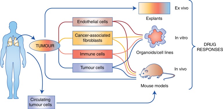
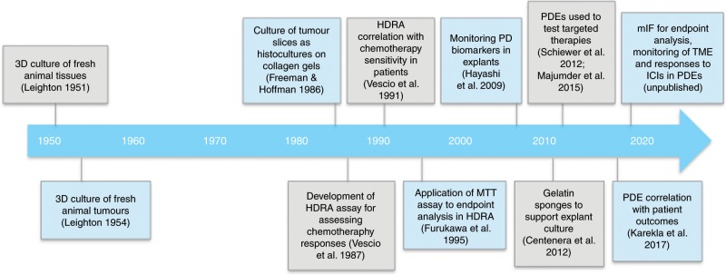
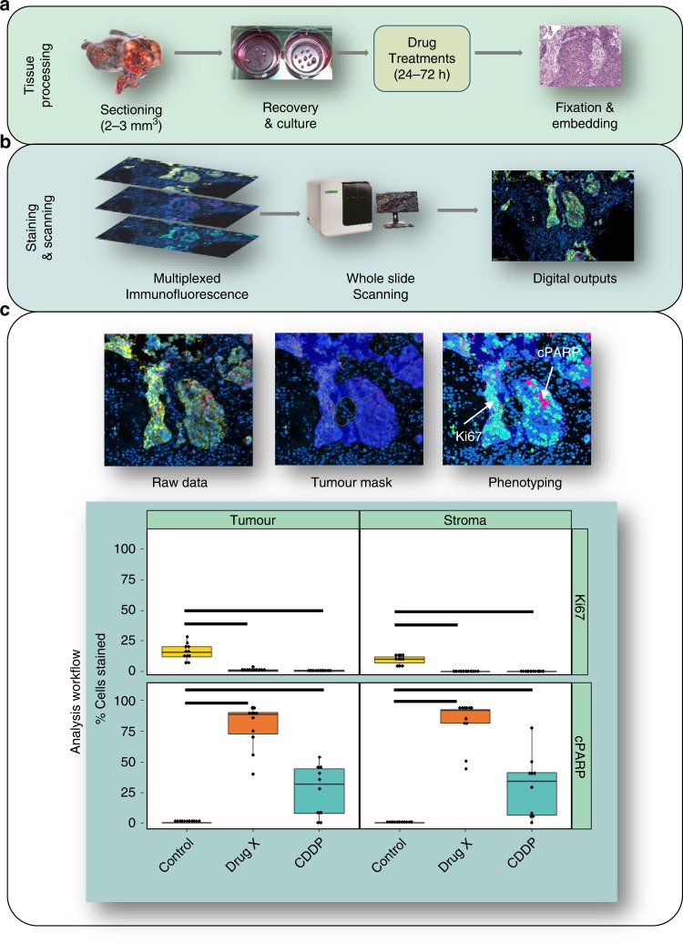
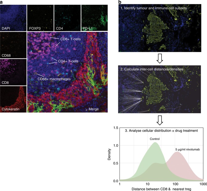
Preclinical models that can accurately predict outcomes in the clinic are much sought after in the field of cancer drug discovery and development. Existing models such as organoids and patient-derived xenografts have many advantages, but they suffer from the drawback of not contextually preserving human tumour architecture. This is a particular problem for the preclinical testing of immunotherapies, as these agents require an intact tumour human-specific microenvironment for them to be effective. In this review, we explore the potential of patient-derived explants (PDEs) for fulfilling this need. PDEs involve the ex vivo culture of fragments of freshly resected human tumours that retain the histological features of original tumours. PDE methodology for anti-cancer drug testing has been in existence for many years, but the platform has not been widely adopted in translational research facilities, despite strong evidence for its clinical predictivity. By modifying PDE endpoint analysis to include the spatial profiling of key biomarkers by using multispectral imaging, we argue that PDEs offer many advantages, including the ability to correlate drug responses with tumour pathology, tumour heterogeneity and changes in the tumour microenvironment. As such, PDEs are a powerful model of choice for cancer drug and biomarker discovery programmes.


Conflict of interest statement

The authors declare no competing interests.

Figures

Fig. 1
Fig. 1. Available preclinical cancer models.
In vitro and in vivo approaches generally involve deconstruction of the original tumours, and in some cases, reconstruction for subsequent assessment of drug responses, although patient-derived xenograft (PDX) models can preserve the integrity of original tumours following immediate transfer. Ex vivo approaches such as patient-derived explant (PDE) models assess drug responses directly in tumour samples obtained fresh from surgery without deconstruction/reconstruction. The schematic shows the cell types available for derivation and use in each model system.
Fig. 2
Fig. 2. Timeline indicating the historical development of patient-derived explant cultures.
HDRA histoculture drug response assay, PD pharmacodynamic, MTT 3-(4,5-dimethylthiazol-2-yl)-2,5-diphenyltetrazolium bromide, PDEs patient-derived explants, mIF multi-immunofluorescence, TME tumour microenvironment, ICI immune-checkpoint inhibitor.
Fig. 3
Fig. 3. Workflow for PDE culture showing multiplexed immunofluorescence outputs and assessment of drug responses in PDEs.
a shows the method for tissue processing, b shows the staining and scanning method and c shows the analysis workflow. In c, the image on the top left shows merged multi-immunofluorescence (mIF) staining of a non-small-cell lung cancer (NSCLC) explant with Ki67, cPARP, pan-cytokeratin and DAPI. The application of the tumour mask (middle) and digitisation of the image (right) allows segregation of staining in the tumour and stroma. The graphs at the bottom depict four quadrants showing % proliferation (Ki67) and % cell death (cPARP) in the stroma and tumour for the NSCLC PDEs treated with vehicle control, cisplatin (CDDP) or experimental Drug X. The PDEs were more responsive to Drug X when compared with cisplatin in both tumour and stroma tissue. Each point represents one PDE with boxplots displaying the first and third quartile (hinges), and median (centre line) with error bars representing the range no further than 1.5× IQR (interquartile range). Significance bars indicate P < 0.05 according to the Kruskal–Wallis test. The findings in this Figure are the authors’ unpublished original data.
Fig. 4
Fig. 4. Multiplexed immunofluorescence.
a A section of a PDE stained with DAPI as well as antibodies specific for immune cell markers (CD4, CD8, CD68 and FOXP3), PD-L1 and pan-cytokeratin followed by TSA-based fluorescent labelling is shown. This method allows for the characterisation of immune-cell subsets in the tumour microenvironment. b Measurement of inter-cell distances in PDEs pre- and post drug treatment. In this example, a melanoma PDE is analysed pre- and post-treatment with the anti-PD1 immunotherapy Nivolumab. To analyse inter-cell distances, PDEs are first stained with relevant cell markers such as tumour or immune-cell subsets followed by digital scanning of images (1). Cells are then identified and phenotyped using Inform software, and distances calculated using the R programming environment (2). The example shown in the histogram (3), indicates increased distance between CD8+ effector T cells and CD4+FOXP3+ regulatory T cells (Tregs) following Nivolumab treatment, confirming on-target effects of this anti-PD1 immunotherapy within the PDE. These images are the authors’ unpublished original data.

References

    1. DiMasi JA, Grabowski HG, Hansen RW. The cost of drug development. N. Engl. J. Med. 2015;372:1972. doi: 10.1056/NEJMc1504317. - DOI - PubMed
    1. Seruga B, Ocana A, Amir E, Tannock IF. Failures in phase III: causes and consequences. Clin. Cancer Res. 2015;21:4552–4560. doi: 10.1158/1078-0432.CCR-15-0124. - DOI - PubMed
    1. Kola I, Landis J. Can the pharmaceutical industry reduce attrition rates? Nat. Rev. Drug Discov. 2004;3:711–715. doi: 10.1038/nrd1470. - DOI - PubMed
    1. Moreno L, Pearson AD. How can attrition rates be reduced in cancer drug discovery? Expert Opin. Drug Discov. 2013;8:363–368. doi: 10.1517/17460441.2013.768984. - DOI - PubMed
    1. Paul SM, Mytelka DS, Dunwiddie CT, Persinger CC, Munos BH, Lindborg SR, et al. How to improve R&D productivity: the pharmaceutical industry’s grand challenge. Nat. Rev. Drug Discov. 2010;9:203–214. doi: 10.1038/nrd3078. - DOI - PubMed

Publication types

Substances