. 2020 Feb;43(2):427-436.
doi: 10.3892/or.2019.7434. Epub 2019 Dec 13.

Dexamethasone induces aberrant macrophage immune function and apoptosis

Affiliations

Dexamethasone induces aberrant macrophage immune function and apoptosis

Fulu Ai et al. Oncol Rep. 2020 Feb.

Abstract

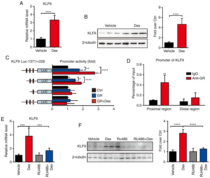
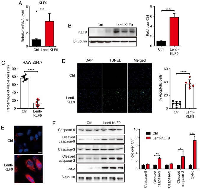
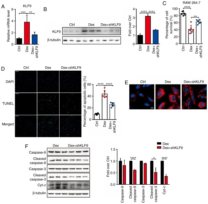
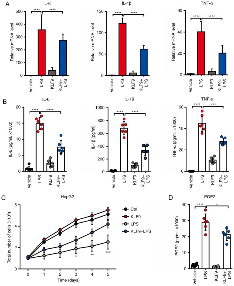
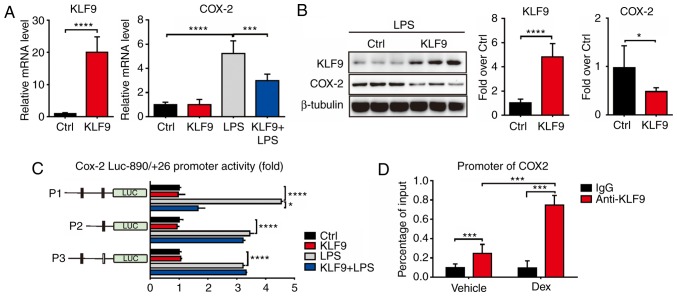
Glucocorticoids (GCs) are known potent clinical drugs, however, their mode of action is still complex and debatable. Macrophages are the most important target of GCs and play a key role in tumor immunity in vivo, but their relationship is also controversial. In the present study, the lentivirus system was used to overexpress and knock down the level of transcription factor Krüppel‑like factor 9 (KLF9). The results revealed that dexamethasone (Dex) induced ROS generation and mitochondria‑dependent apoptosis in RAW 264.7 cells via the KLF9. In addition, overexpression of KLF9 significantly increased apoptosis of RAW 264.7 cells. Notably, ELISA assay revealed that increased expression of KLF9 inhibited LPS‑induced COX‑2 expression and reduced COX‑2‑derived prostaglandin E2 and pro‑inflammatory cytokine secretion. Furthermore, a co‑culture system was used to reveal that overexpression of KLF9 in RAW 264.7 cells promoted HepG2 cell survival. In summary, it is reported that KLF9 promoted apoptosis of proinflammatory macrophages, and suppressed the antitumor effects, which can be selectively targeted by GCs as a novel mechanism to suppress antineoplastic activity.

Keywords: glucocorticoids; macrophages; dexamethasone; KLF9; apoptosis; immunosuppression; COX-2.


Figures

Figure 1.
Figure 1.
Expression of KLF9 is increased by Dex in RAW264.7 cells. (A) Real-time quantitative PCR analysis of the mRNA level of KLF9 in RAW264.7 cells 16 h after treatment with saline or Dex (100 nM) (n=5). (B) Western blot analysis of KLF9 in RAW264.7 cells as described in A; quantitative data are on the right. (C) A series of promoter activity of KLF9 was detected in RAW264.7 cells with saline or Dex (100 nM) treatment. (D) A ChIP assay was performed using anti-GR antibody in RAW264.7 cells. (E) Real-time quantitative PCR analysis of KLF9 in RAW264.7 cells treated with 100 nM Dex or with 10 µM of the GR antagonist RU486 for 16 h. (F) Western blot analysis of the protein level of KLF9 described in E; quantitative data are on the right. Experiments were performed in triplicate. Error bars represent the standard deviation *P<0.05; **P<0.01; ***P<0.005; ****P<0.001. KLF9, Krüppel-like factor 9; Dex, dexamethasone; ChIP, chromatin immunoprecipitation; GR, glucocorticoid receptor.
Figure 2.
Figure 2.
KLF9 increases apoptosis of RAW264.7 cells and activates the mitochondrial apoptotic pathway. (A) Real-time quantitative PCR analysis of the mRNA level of KLF9 in RAW264.7 cells 48 h after infection with control or KLF9 lentivirus (n=5). (B) Western blot analysis of the protein level of KLF9 in RAW264.7 cells as described in A; quantitative data are on the right. (C) Quantitative data of viable cells assessed by the Trypan blue exclusion method. (D) TUNEL assay revealed apoptosis of RAW264.7 cells in KLF9 and the control groups. Quantitative data of the percentage of TUNEL-positive cells is presented on the right. (E) Mitochondrial ROS generation in RAW264.7 cells were analyzed by immunofluorescence (red). Nuclei were stained with DAPI (blue). (F) Western blot analysis was performed for the detection of the mitochondrial-dependent apoptotic pathway including procaspase-9, cleaved caspase-9, procaspase-3, cleaved caspase-3 and Cyt-c proteins, quantitative data are on the right. Experiments were performed in triplicate. Error bars represent the standard deviation (*P<0.05; **P<0.01; ***P<0.005; ****P<0.001). KLF9, Krüppel-like factor 9; Dex, dexamethasone; ROS, reactive oxygen species; Cyt-c, cytochrome c.
Figure 3.
Figure 3.
Knockdown of KLF9 decreases apoptosis and inhibits the activation of the mitochondrial apoptotic pathway induced by Dex in RAW264.7 cells. (A) Real-time quantitative PCR analysis of the mRNA level of KLF9 in RAW264.7 cells treated with Dex or saline after infection with control or KLF9 lentivirus for 48 h. (B) Western blot analysis of the protein level of KLF9 in RAW264.7 cells as described in A. (C) Viable cells were analyzed using the Trypan blue exclusion method. (D) A TUNEL assay revealed the apoptosis of RAW264.7 cells in different groups. Quantitative data of TUNEL-positive cell percentages are on the right. (E) Mitochondrial ROS generation in RAW264.7 cells were analyzed by immunofluorescence. Nuclei were stained with DAPI (blue). (F) Western blot analysis was performed for the detection of the mitochondrial-dependent apoptotic pathway including procaspase-9, cleaved caspase-9, procaspase-3, cleaved caspase-3 and Cyt-c proteins; quantitative data are on the right. Experiments were performed in triplicate. Error bars represent the standard deviation (*P<0.05; **P<0.01; ***P<0.005; ****P<0.001). KLF9, Krüppel-like factor 9; Dex, dexamethasone; ROS, reactive oxygen species; Cyt-c, cytochrome c.
Figure 4.
Figure 4.
Effects of KLF9 on the production of proinflammatory cytokines from LPS-induced RAW264.7 cells. (A) RAW264.7 cells were transfected with Lenti-KLF9 and then co-treated with 1 µg/ml LPS for 24 h. The mRNA levels of TNF-α, IL-1β, IL-6 are analyzed by Real-time quantitative PCR analysis. (B) An ELISA assay was used to determine TNF-α, IL-1β, IL-6 production in the supernatants of RAW264.7 cells following treatment with 1 µg/ml LPS and transfected with Lenti-KLF9 or control, respectively. (C) Proliferation rate of HepG2 cells was assessed using CCK8 after co-culture with RAW264.7 cells following treatment with 1 µg/ml LPS and transfected with Lenti-KLF9 or control, respectively. (D) ELISA assay was used to determine PGE2 production in the supernatants of RAW264.7 cells following treatment with 1 µg/ml LPS and transfected with Lenti-KLF9 or control, respectively. Experiments were performed in triplicate. Error bars represent the standard deviation (*P<0.05; **P<0.01; ***P<0.005; ****P<0.001). KLF9, Krüppel-like factor 9; LPS, lipopolysaccharide.
Figure 5.
Figure 5.
KLF9 suppresses LPS-induced COX-2 expression. (A and B) Real-time quantitative PCR and western blot analysis revealed the expression levels of COX-2 with KLF9 overexpression and control RAW264.7 cells following treatment with 1 µg/ml LPS or saline. (C) The promoter region of COX-2 was cloned and fused to a luciferase reporter system. The deleted and mutated promoters were co-transfected into RAW264.7 cells along with pcDNA3.1 or KLF9 expression plasmid. Luciferase activity was corrected for Renilla luciferase activity and normalized to control groups. (D) ChIP analysis in RAW264.7 cells following LPS treatment. Experiments were performed in triplicate. Error bars represent the standard deviation (*P<0.05; ***P<0.005; ****P<0.001). KLF9, Krüppel-like factor 9; COX-2, cyclooxygenase-2; ChIP, chromatin immunoprecipitation; LPS, lipopolysaccharide.

References

    1. Cari L, De Rosa F, Nocentini G, Riccardi C. Context-dependent effect of glucocorticoids on the proliferation, differentiation, and apoptosis of regulatory T cells: A review of the empirical evidence and clinical applications. Int J Mol Sci. 2019;20(pii):E1142. doi: 10.3390/ijms20051142. - DOI - PMC - PubMed
    1. Mylka V, Deckers J, Ratman D, De Cauwer L, Thommis J, De Rycke R, Impens F, Libert C, Tavernier J, Vanden Berghe W, et al. The autophagy receptor SQSTM1/p62 mediates anti-inflammatory actions of the selective NR3C1/glucocorticoid receptor modulator compound A (CpdA) in macrophages. Autophagy. 2018;14:2049–2064. doi: 10.1080/15548627.2018.1495681. - DOI - PMC - PubMed
    1. Patil RH, Naveen Kumar M, Kiran Kumar KM, Nagesh R, Kavya K, Babu RL, Ramesh GT, Chidananda Sharma S. Dexamethasone inhibits inflammatory response via down regulation of AP-1 transcription factor in human lung epithelial cells. Gene. 2018;645:85–94. doi: 10.1016/j.gene.2017.12.024. - DOI - PubMed
    1. Achuthan A, Aslam ASM, Nguyen Q, Lam PY, Fleetwood AJ, Frye AT, Louis C, Lee MC, Smith JE, Cook AD, et al. Glucocorticoids promote apoptosis of proinflammatory monocytes by inhibiting ERK activity. Cell Death Dis. 2018;9:267. doi: 10.1038/s41419-018-0332-4. - DOI - PMC - PubMed
    1. Li B, Wang Y, Yin L, Huang G, Xu Y, Su J, Ma L, Lu J. Glucocorticoids promote the development of azoxymethane and dextran sulfate sodium-induced colorectal carcinoma in mice. BMC Cancer. 2019;19:94. doi: 10.1186/s12885-019-5299-8. - DOI - PMC - PubMed