. 2020 Feb;45(2):556-568.
doi: 10.3892/ijmm.2019.4447. Epub 2019 Dec 27.

Protective effects of human umbilical cord blood‑derived mesenchymal stem cells against dexamethasone‑induced apoptotic cell death in hair follicles

Affiliations

Protective effects of human umbilical cord blood‑derived mesenchymal stem cells against dexamethasone‑induced apoptotic cell death in hair follicles

Dong Ho Bak et al. Int J Mol Med. 2020 Feb.

Abstract

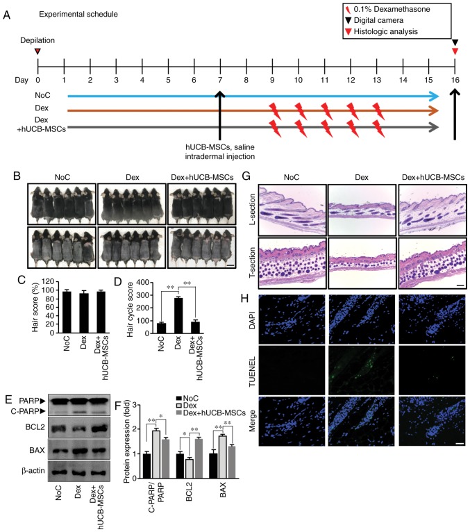
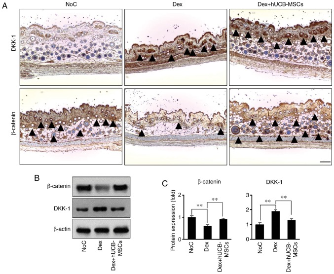
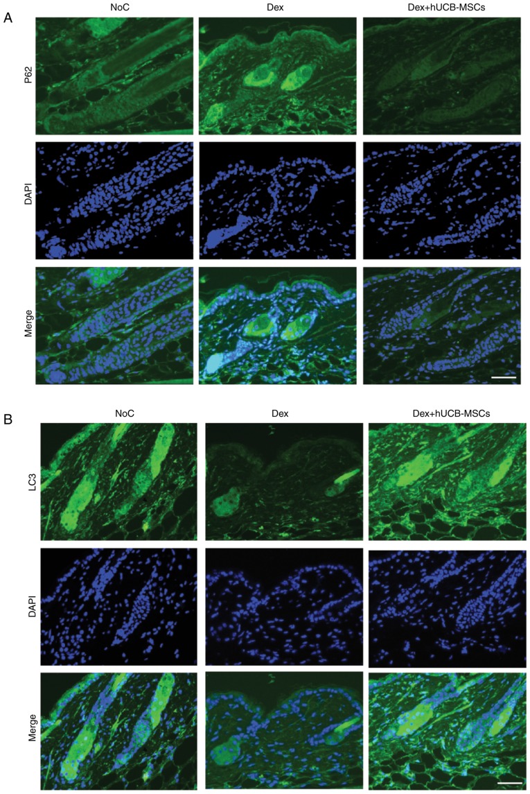
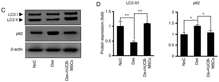
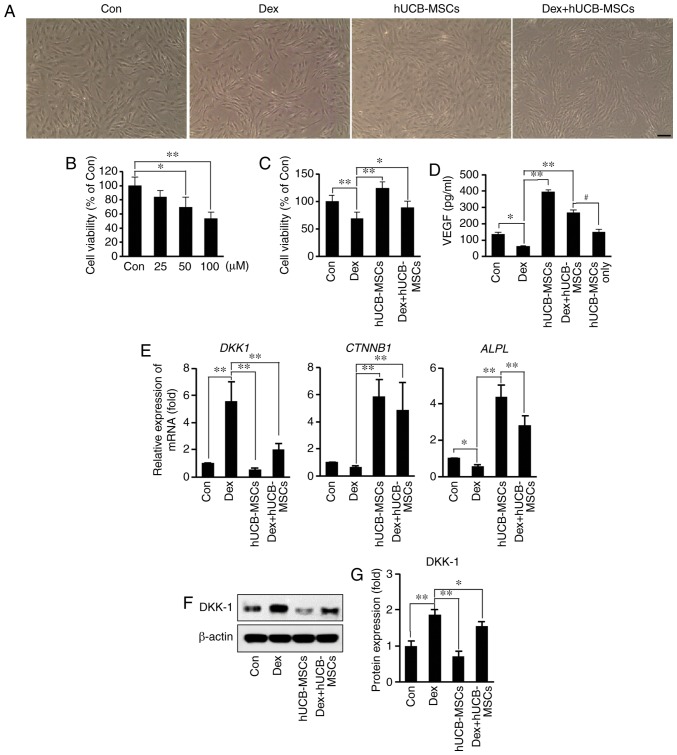
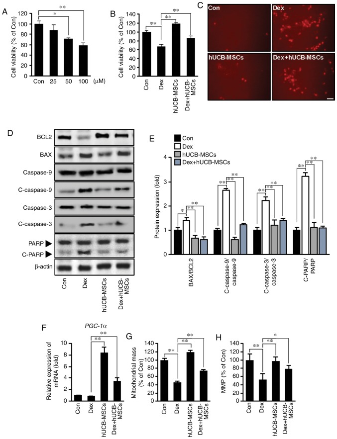
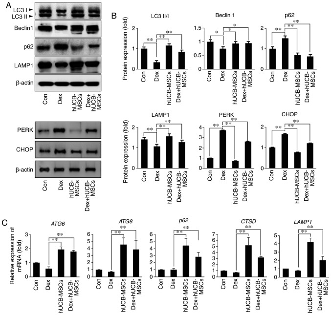
Alopecia is a common and distressing condition, and developing new therapeutic agents to prevent hair loss is important. Human umbilical cord blood‑derived mesenchymal stem cells (hUCB‑MSCs) have been studied intensively in regenerative medicine. However, the therapeutic potential of these cells against hair loss and hair organ damage remains unclear, and the effects of hUCB‑MSC transplantation on hair loss require evaluation. The current study aimed to investigate the effects of hUCB‑MSCs on hair regression in vivo and restoration of anagen conduction on hair growth in vitro. The effects of hUCB‑MSCs were explored in mouse catagen induction models using a topical treatment of 0.1% dexamethasone to induce hair regression. Dexamethasone was also used to simulate a stress environment in vitro. The results demonstrated that hUCB‑MSCs significantly prevented hair regression induced by dexamethasone topical stimulation in vivo. Additionally, hUCB‑MSCs significantly increased the proliferation of human dermal papilla cells (hDPCs) and HaCaT cells, which are key constituent cells of the hair follicle. Stimulation of vascular endothelial growth factor secretion and decreased expression of DKK‑1 by hUCB‑MSCs were also observed in hDPCs. Restoration of cell viability by hUCB‑MSCs suggested that these cells exerted a protective effect on glucocorticoid stress‑associated hair loss. In addition, anti‑apoptotic effects and regulation of the autophagic flux recovery were observed in HaCaT cells. The results of the present study indicated that hUCB‑MSCs may have the capacity to protect hair follicular dermal papilla cells and keratinocytes, thus preventing hair loss. Additionally, the protective effects of hUCB‑MSCs may be resistant to dysregulation of autophagy under harmful stress.

Keywords: hair follicle; hUcB-MScs; alopecia; stem-cell therapy; glucocorticoid.


Figures

Figure 1
Figure 1
hUCB-MSCs prevent the catagen phase in C57BL/6 mice. (A) Experimental schedule. On day 7 of tape depilation, the NoC group was treated with 800 µl saline; 1.25×10⁵ cells/ml of hUCB-MSCs were injected into the intradermal site in the Dex + hUCB-MSCs group, and the Dex group was not treated. From day 9 to 13, 1 ml dexamethasone (0.1%) was topically administered to the Dex and Dex + hUCB-MSCs groups. On day 16, all animals were sacrificed. (B) The dorsal skin was photographed on day 16 (upper panel, pre-depilation; lower panel, post-depilation). Scale bar, 2.5 cm. (C) Quantification of the hair score. Data are expressed as the mean ± SD. (D) HCS on day 16 post-depilation. Higher HCS indicates the progression of catagen. (E) Western blot analysis of PARP, Bcl-2 and Bax levels in dorsal tissue from the three groups. (F) Densitometry analysis if protein expression. (G) Sections of the dorsal skin. L-section, longitudinal section; T-section, transverse section. Scale bar, 100 µm. (H) TUNEL staining. Representative images of TUNEL staining (green fluorescence, middle panel). DAPI was used as a counterstain (upper panel), and merged pictures are presented in the bottom panel. Scale bar, 25 µm. *P<0.05, **P<0.01 vs. Dex. NoC, normal control; Dex, dexamethasone; HCS, hair cycle score; hUCB-MSCs, human umbilical cord blood-derived mesenchymal stem cells; PARP, poly (ADP-ribose) polymerase.
Figure 2
Figure 2
Effects of hUCB-MSCs on DKK-1 and β-catenin expression in mouse skin hair follicles. (A) Representative images of immunohistochemistry. Protein location was observed using anti-DKK-1 (upper panel) and anti-β-catenin (lower panel) antibodies. Black arrow heads indicate a positive reaction. Scale bar, 100 µm. (B) Western blot analysis of DKK-1 and β-catenin levels in dorsal skin tissue in the three groups. (C) Bar diagram of densitometry analysis. **P<0.01 vs. Dex. NoC, normal control; Dex, dexamethasone; hUCB-MSCs, human umbilical cord blood-derived mesenchymal stem cells; DKK-1, Dickkopf WNT signaling pathway inhibitor 1.
Figure 3
Figure 3
Effects of hUCB-MSCs on LC3 and p62 expression in mouse skin hair follicles. (A and B) Representative immunofluorescence images. Protein localization was observed using (A) anti-p62 and (B) anti-LC3 antibodies. DAPI was used as a counterstain. Scale bar, 100 µm. (C) Western blot analysis of LC3 and p62 levels in dorsal skin tissues of the 3 groups. (D) Bar diagram of densitometry analysis. *P<0.05, **P<0.01 vs. Dex. NoC, normal control; Dex, dexamethasone; hUCB-MSCs, human umbilical cord blood-derived mesenchymal stem cells; LC3, microtubule-associated protein 1 light chain 3 β.
Figure 3
Figure 3
Effects of hUCB-MSCs on LC3 and p62 expression in mouse skin hair follicles. (A and B) Representative immunofluorescence images. Protein localization was observed using (A) anti-p62 and (B) anti-LC3 antibodies. DAPI was used as a counterstain. Scale bar, 100 µm. (C) Western blot analysis of LC3 and p62 levels in dorsal skin tissues of the 3 groups. (D) Bar diagram of densitometry analysis. *P<0.05, **P<0.01 vs. Dex. NoC, normal control; Dex, dexamethasone; hUCB-MSCs, human umbilical cord blood-derived mesenchymal stem cells; LC3, microtubule-associated protein 1 light chain 3 β.
Figure 4
Figure 4
Effects of hUCB-MSC co-culture on viability, VEGF secretion, mRNA and protein expression in hDPCs following Dex stimulation. (A) Representative images of hDPCs. Cell density was decreased by Dex stimulation; hUCB-MSCs counteracted the reduction. Scale bar, 50 µm. (B) hDPCs were treated with 25-100 µM Dex for 48 h. Cell viability was determined via MTT assay. *P<0.05, **P<0.01 vs. Con. (C) Effects of hUCB-MSCs on the viability of hDPCs stimulated with 100 µM Dex for 48 h in the presence or absence of hUCB-MSCs. Cell viability was determined via MTT assay. *P<0.05, **P<0.01 vs. Dex. (D) Secretory concentration of VEGF. hDPCs were stimulated with 100 µM Dex for 48 h in the presence or absence of hUCB-MSCs, VEGF concentration in the culture supernatant was evaluated using ELISA. *P<0.05, **P<0.01 vs. Dex,; #P<0.05 vs. Dex + hUCB-MSCs. (E) Expression of DKK-1, CTNNB1 and ALPL mRNA was determined by reverse transcription-quantitative PCR in hDPCs stimulated with 100 µM Dex for 24 h in the presence or absence of hUCB-MSCs. *P<0.05, **P<0.01 vs. Dex. (F) Western blot analysis of DKK-1 levels in hDPCs stimulated with 100 µM Dex for 24 h in the presence or absence of hUCB-MSCs. (G) Bar diagram of densitometry analysis. *P<0.05, **P<0.01 vs. Dex. hDPCs, human dermal papilla cells; Con, control; Dex, dexamethasone; hUCB-MSC, human umbilical cord blood-derived mesenchymal stem cell; VEGF, vascular endothelial growth factor; DKK-1, Dickkopf WNT signaling pathway inhibitor 1; CTNNB1, β-catenin; ALPL, human alkaline phosphatase liver/bone/kidney isozyme.
Figure 5
Figure 5
Effects of hUCB-MSC co-culture on apoptosis and mitochondria biogenesis after Dex stimulation in HaCaT cells. (A) HaCaT cells were treated with 25-100 µM Dex for 48 h. Cell viability was determined using MTT assay. *P<0.05, **P<0.01 vs. Con. (B) Effects of hUCB-MSCs on the viability of HaCaT cells stimulated with 100 µM Dex for 48 h in the presence or absence of hUCB-MSCs. Cell viability was determined via MTT assay. **P<0.01 vs. Dex. (C) Representative images of HaCaT cells stained with PI. Positive reactions were increased by Dex stimulation, whereas hUCB-MSCs counteracted apoptotic reactions. Scale bar, 50 µm. (D) Western blot analysis of Bcl-2, Bax, caspase-9, C-caspase-9, caspase-3, C-caspase-3 and PARP levels in HaCaT cells stimulated with 100 µM Dex for 48 h in the presence or absence of hUCB-MSCs. (E) Bar diagram of densitometry analysis. **P<0.01 vs. Dex. (F) PGC-1α mRNA expression was determined using reverse transcription-quantitative PCR in HaCaT cells stimulated with 100 µM Dex for 24 h in the presence or absence of hUCB-MSCs. **P<0.01 vs. Dex. (G) Mitochondrial mass was measured using MitoTracker fluorescence signals. **P<0.01 vs. Dex. (H) MMP was measured under the indicated conditions using the MMP-sensitive probe. *P<0.05, **P<0.01 vs. Dex. Con, control; Dex, dexamethasone; hUCB-MSC, human umbilical cord blood-derived mesenchymal stem cell; PI, propidium iodide; C, cleaved; PARP, poly (ADP-ribose) polymerase; MMP, mitochondrial membrane potential; PGC-1α, peroxisome proliferator-activated receptor gamma coactivator 1α.
Figure 6
Figure 6
Effects of hUCB-MSC co-culture on autophagy and ER stress in HaCaT cells following Dex stimulation. (A) HaCaT cell lysates were immunoblotted with antibodies against LC3I/II, Beclin1, p62, LAMP1, PERK, CHOP or β-actin. (B) Densitometry analysis results. **P<0.01 vs. Dex group. (C) Transcript levels of autophagy-related genes were determined by reverse transcription-quantitative PCR. **P<0.01 vs. Dex group. LC3, microtubule-associated protein 1 light chain 3 β Con, control; Dex, dexamethasone; hUCB-MSC, human umbilical cord blood-derived mesenchymal stem cell; LAMP1, human lysosomal-associated membrane protein 1; CHOP, transcription factor C/EBP homologous protein; PERK, protein kinase R (PKR)-like endoplasmic reticulum kinase; ATG, autophagy-related protein.

References

    1. Ding DC, Chang YH, Shyu WC, Lin SZ. Human umbilical cord mesenchymal stem cells: A new era for stem cell therapy. Cell Transplant. 2015;24:339–347. doi: 10.3727/096368915X686841. - DOI - PubMed
    1. Oh W, Kim DS, Yang YS, Lee JK. Immunological properties of umbilical cord blood-derived mesenchymal stromal cells. Cell Immunol. 2008;251:116–123. doi: 10.1016/j.cellimm.2008.04.003. - DOI - PubMed
    1. Chen M, Xiang Z, Cai J. The anti-apoptotic and neuro-protective effects of human umbilical cord blood mesenchymal stem cells (hUCB-MSCs) on acute optic nerve injury is transient. Brain Res. 2013;1532:63–75. doi: 10.1016/j.brainres.2013.07.037. - DOI - PubMed
    1. Kang SG, Jeun SS, Lim JY, Kim SM, Yang YS, Oh WI, Huh PW, Park CK. Cytotoxicity of human umbilical cord blood-derived mesenchymal stem cells against human malignant glioma cells. Child's Nerv Syst. 2008;24:293–302. doi: 10.1007/s00381-007-0515-2. - DOI - PubMed
    1. Lim JY, Jeong CH, Jun JA, Kim SM, Ryu CH, Hou Y, Oh W, Chang JW, Jeun SS. Therapeutic effects of human umbilical cord blood-derived mesenchymal stem cells after intrathecal administration by lumbar puncture in a rat model of cerebral ischemia. Stem Cell Res Ther. 2011;2:38. doi: 10.1186/scrt79. - DOI - PMC - PubMed