. 2020 Nov;77(21):4413-4428.
doi: 10.1007/s00018-019-03425-6. Epub 2020 Jan 2.

hnRNPA2B1 inhibits the exosomal export of miR-503 in endothelial cells

Affiliations

hnRNPA2B1 inhibits the exosomal export of miR-503 in endothelial cells

Jennifer Pérez-Boza et al. Cell Mol Life Sci. 2020 Nov.

Abstract

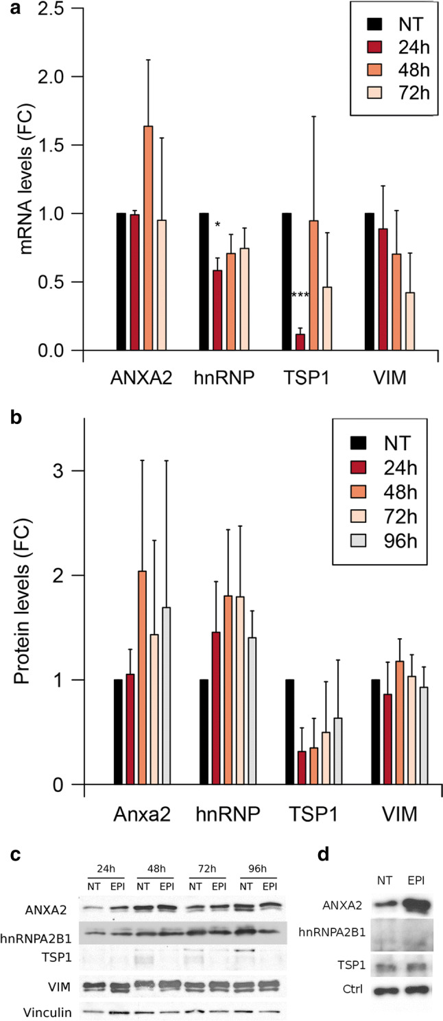
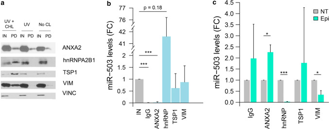
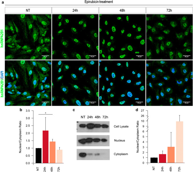
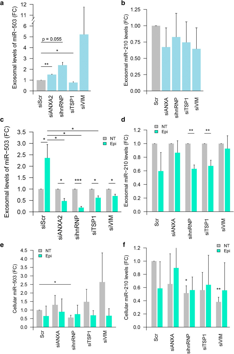
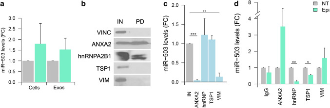
The chemotherapeutic drug epirubicin increases the exosomal export of miR-503 in endothelial cells. To understand the mechanisms behind this process, we transfected endothelial cells with miR-503 carrying a biotin tag. Then, we pulled-down the proteins interacting with miR-503 and studied their role in microRNA exosomal export. A total of four different binding partners were identified by mass spectrometry and validated by western blotting and negative controls, among them ANXA2 and hnRNPA2B1. Using knock-down systems combined with pull-down analysis, we determined that epirubicin mediates the export of miR-503 by disrupting the interaction between hnRNPA2B1 and miR-503. Then, both ANXA2 and miR-503 are sorted into exosomes while hnRNPA2B1 is relocated into the nucleus. The combination of these processes culminates in the increased export of miR-503. These results suggest, for the first time, that RNA-binding proteins can negatively regulate the exosomal sorting of microRNAs.

Keywords: EVs; Exosomal export; Exosomes; MicroRNAs; RNA-binding proteins.


Conflict of interest statement

The authors declare no competing interests.

Figures

Fig. 1
Fig. 1
Exosome characterization and identification of miR-503 binding proteins: a exosome characterization of HUVEC lysates and exosomes (10 µg) against the EV markers: CD9, CD63, CD81, syntenin (SYN), and the cellular marker cytochrome C (Cyt C). b Dynamic light scattering analysis of exosomal preparations. c Electron microscopy images of HUVEC exosomes labeled with anti-CD63 and anti-CD105, scale bars = 100 nm. d Cells were treated with epirubicin for 24 h. Cell lysates were prepared 24 h after treatment and exosomes were collected 72 h after removing the chemotherapeutic drug. Cellular and exosomal levels of miR-503 were evaluated by qPCR. Data show mean ± SEM (n = 3). **p < 0.01 vs. respective control. e Schematic representation of the protocol used to identify the MEC proteins. HUVECs (30 × 106 cells) were transfected with miR-503-biotin (10 nM). The following day, the cells were crosslinked with DTSSP and UV. HUVECs lysates were incubated with streptavidin beads. Both input (IN) (cellular lysate) and pull-down fractions (PD) were separated by SDS-PAGE. Isolated proteins were identified by mass spectrometry and f validated by western blotting against pull-down (PD) and input (IN) (1% of cell lysate) fractions using vinculin as loading control. Cel-miR-67 was used as a negative control for the pull-down
Fig. 2
Fig. 2
Epirubicin regulates the mRNA levels of TSP1 and hnRNPA2B1. HUVECs were treated with epirubicin for 24 h and the levels of a RNA and b proteins were assessed, respectively, by qPCR at 24, 48, and 72 h after treatment and western blotting at 24, 48, 72, and 96 h after treatment. Data represents fold change against non-treated cells. Plots show mean and SEM from three independent experiments. *p < 0.05, ***p < 0.001. c Representative western blot. d HUVECs were treated with epirubicin for 24 h and exosomes were produced for additional 72 h. Exosomes were purified by ultracentrifugation and the levels of proteins were determined by western blotting (10 µg/lane). Representative example of N = 3
Fig. 3
Fig. 3
Epirubicin disrupts the interaction between miR-503 and VIM and hnRNPA2B1. a HUVECs (30·106 cells) were transfected with miR-503-biotin (10 nM). The following day, the cells were crosslinked with DTSSP and UV (UV + CHL), only UV (UV) or not subjected to any crosslinking (No CL). HUVECs lysates were incubated with streptavidin beads. Both input (IN) (cellular lysate) and pull-down fractions (PD) were separated by SDS-PAGE and revealed by western blotting using indicated proteins. Input (IN) = 1% of cell lysate. Vinculin was used as loading control. b, c HUVECs were transfected with miR-503 (10 nM). 48 h later, immunoprecipitation assays were performed using the indicated antibodies or an IgG control and the levels of miR-503 were evaluated by qPCR. Plots show b fold change of miR-503 in the immunoprecipitated (IP) vs input fractions (IN) in non-treated cells and c fold change of immunoprecipitated miR-503 in epirubicin-treated (EPI) vs non-treated cells (NT). Plots represent mean and SEM from three independent experiments,*p < 0.05; **p < 0.01, ***p < 0.001. IN = 1% of the cellular lysate before immunoprecipitation
Fig. 4
Fig. 4
hnRNPA2B1 re-localizes into the nucleus after epirubicin treatment. a Confocal images of hnRNPA2B1 (green) and DAPI (blue) of HUVECs treated with epirubicin for 24 h. Pictures taken at the indicated times after starting the treatment. b Quantification of the intensity in the cytoplasm and nucleus were performed using ImageJ on epifluorescence images. c Nuclear and cytoplasmic fractions from HUVECs treated with epirubicin and analysed by western blotting for hnRNPA2B1 localization. d Abundance ratio of hnRNPA2B1 between nucleus and cytoplasm in subcellular fractions. All plots show results from three independent experiments. *p < 0.05. Validation of subcellular fractionation method in Supplementary Figure S4
Fig. 5
Fig. 5
ANXA2, hnRNPA2B1, TSP1 and VIM are necessary for the effect of epirubicin on the exosomal export of miR-503. HUVECs were transfected with siRNA for the indicated protein or control siRNA (siScr) (20 nM). Then, cells were treated with epirubicin for 24 h (or not) and exosomes were produced for the following 72 h. The levels of miR-503 (a, c, e) and miR-210 (b, d, f) in purified cells and exosomes were then assessed via qPCR. Data show fold change of miR-503 (a) and miR-210 (b) in exosomes from siRNA-transfected cells vs Scramble RNA-transfected (siScr) cells in non-treated conditions. Fold change of miR-503 and mR-210 respectively in exosomes (c, d) and cells (e, f) after siRNA transfection and epirubicin treatment normalized to untreated conditions. Plots show mean and SEM of three independent experiments. (*p < 0.05; **p < 0.01, ***p < 0.001)
Fig. 6
Fig. 6
ANXA2 and hnRNPA2B1 are necessary for the effect of epirubicin on the exosomal export of miR-503 in microvascular endothelial cells. HMVECs were treated with epirubicin for 24 h. Cell lysates were prepared 24 h after treatment and exosomes were collected 72 h after removing the chemotherapeutic drug. Cellular and exosomal levels of miR-503 were evaluated by qPCR. Plot represents mean and SEM (n = 3). b HMVECs were transfected with miR-503-biotin (10 nM). The following day, the cells were crosslinked with DTSSP and UV. HMECs lysates were incubated with streptavidin beads. Both input (IN) (cellular lysate) and pull-down fractions (PD) were separated by SDS-PAGE. Previously identified components of the MEC complex were validated by western blotting against pull-down (PD) and input (IN) (1% of cell lysate) fractions using vinculin as loading control. c HMVECs were transfected with miR-503 (10 nM). 48 h later, immunoprecipitation assays were performed using the indicated antibodies and the levels of miR-503 were evaluated by qPCR. Plot shows fold change of miR-503 in the immunoprecipitated (IP) vs input fractions (IN) in non-treated cells and (d) fold change of immunoprecipitated miR-503 in epirubicin-treated (EPI) vs non-treated cells (NT). Plots represent mean and SEM from three independent experiments, *p < 0.05, **p < 0.01, ***p < 0.001. IN = 1% of the cellular lysate before immunoprecipitation

References

    1. van Niel G, D’Angelo G, Raposo G. Shedding light on the cell biology of extracellular vesicles. Nat Rev Mol Cell Biol. 2018 doi: 10.1038/nrm.2017.125. - DOI - PubMed
    1. McKelvey KJ, Powell KL, Ashton AW, et al. Exosomes: mechanisms of uptake. J Circ Biomark. 2015 doi: 10.5772/61186. - DOI - PMC - PubMed
    1. Dreyer F, Baur A. Biogenesis and functions of exosomes and extracellular vesicles. Methods Mol Biol. 2016;1448:201–216. doi: 10.1007/978-1-4939-3753-0_15. - DOI - PubMed
    1. Pérez-Boza J, Lion M, Struman I. Exploring the RNA landscape of endothelial exosomes. RNA. 2018;24:423–435. doi: 10.1261/rna.064352.117. - DOI - PMC - PubMed
    1. Kalluri R, LeBleu VS (2017) Discovery of double-stranded genomic DNA in circulating exosomes. Cold Spring Harb Symp Quant Biol LXXXI:030932. 10.1101/sqb.2016.81.030932 - DOI - PubMed

MeSH terms