

Current advances in biodegradable synthetic polymer based cardiac patches
- PMID: 31895482
- PMCID: PMC7030941
- DOI: 10.1002/jbm.a.36874
Current advances in biodegradable synthetic polymer based cardiac patches
Abstract
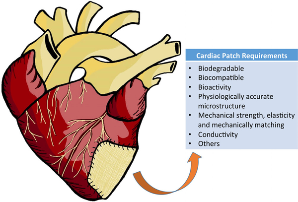
The number of people affected by heart disease such as coronary artery disease and myocardial infarction increases at an alarming rate each year. Currently, the methods to treat these diseases are restricted to lifestyle change, pharmaceuticals, and eventually heart transplant if the condition is severe enough. While these treatment options are the standard for caring for patients who suffer from heart disease, limited regenerative ability of the heart restricts the effectiveness of treatment and may lead to other heart-related health problems in the future. Because of the increasing need for more effective therapeutic technologies for treating diseased heart tissue, cardiac patches are now a large focus for researchers. The cardiac patches are designed to be integrated into the patients' natural tissue to introduce mechanical support and healing to the damaged areas. As a promising alternative, synthetic biodegradable polymer based biomaterials can be easily manipulated to customize material properties, as well as possess certain desired characteristics for cardiac patch use. This comprehensive review summarizes recent works on synthetic biodegradable cardiac patches implanted into infarcted animal models. In addition, this review describes the basic requirements that should be met for cardiac patch development, and discusses the inspirations to designing new biomaterials and technologies for cardiac patches.
Keywords: biodegradable polymer; cardiac patch; heart disease; heart infarction.
© 2020 Wiley Periodicals, Inc.
Figures
References
-
- Alperin C, Zandstra PW, & Woodhouse KA (2005). Polyurethane films seeded with embryonic stem cell-derived cardiomyocytes for use in cardiac tissue engineering applications. Biomaterials, 26(35), 7377–7386. - PubMed
-
- Bahrami S, Solouk A, Mirzadeh H, & Seifalian AM (2019). Electroconductive polyurethane/graphene nanocomposite for biomedical applications. Composites. Part B, Engineering, 168, 421–431.
-
- Benjamin EJ, Virani SS, Callaway CW, Chamberlain AM, Chang AR, Cheng S, … American Heart Association Council on Epidemiology and Prevention Statistics Committee and Stroke Statistics Subcommittee. (2018). Heart disease and stroke statistics-2018 update: A report from the American Heart Association. Circulation, 137(12), e67–e492. - PubMed
-
- Borriello A, Guarino V, Schiavo L, Alvarez-Perez MA, & Ambrosio L (2011). Optimizing PANi doped electroactive substrates as patches for the regeneration of cardiac muscle. Journal of Materials Science. Materials in Medicine, 22(4), 1053–1062. - PubMed
Publication types
MeSH terms
Substances
Grants and funding
LinkOut - more resources
Full Text Sources
Research Materials
Miscellaneous
