. 2020 Jan 2;16(1):e1008473.
doi: 10.1371/journal.pgen.1008473. eCollection 2020 Jan.

DNA double strand break repair in Escherichia coli perturbs cell division and chromosome dynamics

Affiliations

DNA double strand break repair in Escherichia coli perturbs cell division and chromosome dynamics

Martin A White et al. PLoS Genet. .

Abstract

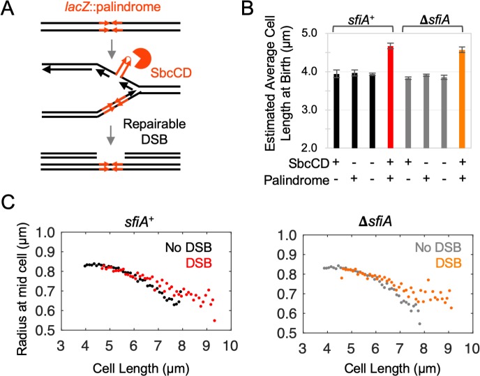
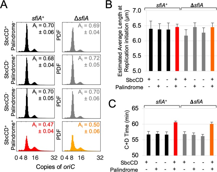
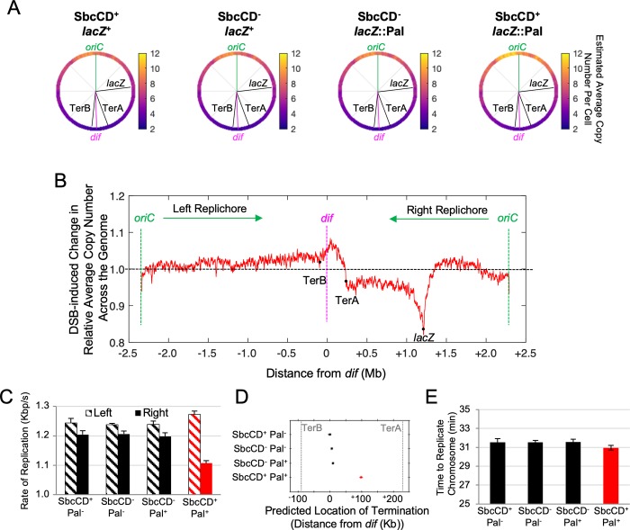
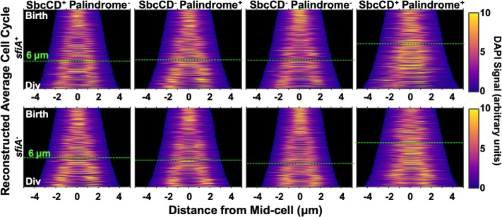
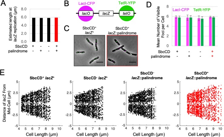
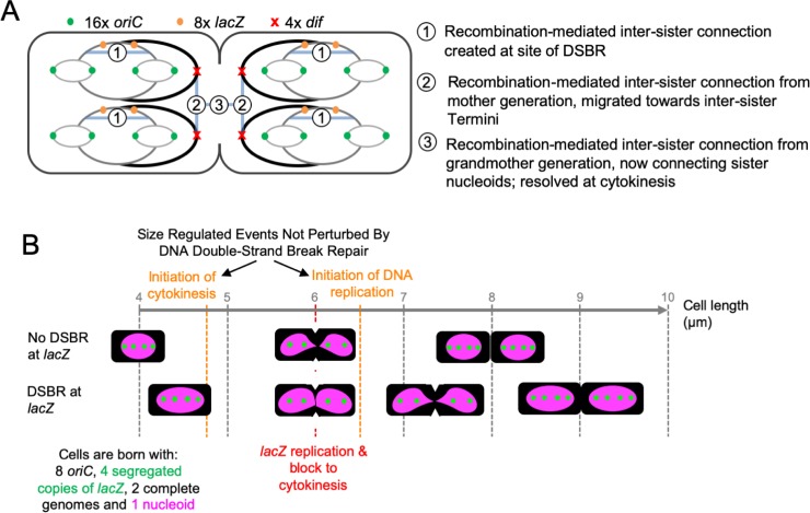
To prevent the transmission of damaged genomic material between generations, cells require a system for accommodating DNA repair within their cell cycles. We have previously shown that Escherichia coli cells subject to a single, repairable site-specific DNA double-strand break (DSB) per DNA replication cycle reach a new average cell length, with a negligible effect on population growth rate. We show here that this new cell size distribution is caused by a DSB repair-dependent delay in completion of cell division. This delay occurs despite unperturbed cell size regulated initiation of both chromosomal DNA replication and cell division. Furthermore, despite DSB repair altering the profile of DNA replication across the genome, the time required to complete chromosomal duplication is invariant. The delay in completion of cell division is accompanied by a DSB repair-dependent delay in individualization of sister nucleoids. We suggest that DSB repair events create inter-sister connections that persist until those chromosomes are separated by a closing septum.


Conflict of interest statement

The authors have declared that no competing interests exist.

Figures

Fig 1
Fig 1. DSBR delays completion of cytokinesis without affecting the size control of its initiation.
A) E. coli cells with an exogenous 246 bp interrupted DNA palindrome integrated into the lacZ gene of their chromosome undergo DNA double-strand break repair (DSBR) by homologous recombination once per replication cycle due to replication-dependent cleavage of the palindrome by the hairpin endonuclease SbcCD. B) Estimated average length at birth derived assuming an idealized population structure of cultures of cells undergoing DSBR (SbcCD+ lacZ::palindrome) and controls; n = 4, error bars show standard error of the mean. C) Radius of mid-cell (the location of cytokinesis) plotted as a function of cell length. Data from four independent cultures were aggregated and mid-cell radii averaged within 100 nm cell length. For both sfiA+ and ΔsfiA strains, the data from the three control strain backgrounds not undergoing DSBR (SbcCD+ lacZ+, SbcCD- lacZ+ and SbcCD- lacZ::palindrome) were averaged for clarity in the plots. No difference was detected between the three control strains in either sfiA+ or ΔsfiA genetic backgrounds.
Fig 2
Fig 2. DSBR does not affect the cell size control of initiation of chromosomal DNA replication.
A) Distribution of the copy number of the single chromosomal origin of replication (oriC) in cells isolated from asynchronous, exponentially growing cultures of E. coli as ascertained by replication runout. Shown are representative results from three independent experiments for each strain. Data are normalized as Probability Distribution Functions (PDFs). Insert of each panel shows the estimated relative cell age at replication initiation (Ai), +/- the standard error of the mean. B) Estimated average cell length at initiation of chromosomal DNA replication calculated using the measured average copy number of oriC from three independent experiments and the estimated average cell length at birth (Fig 1B). C) C+D time (a component of the Cooper-Helmstetter model) calculated using the average mass doubling time for all strains (S1B Fig) and the average cellular oriC copy number for each strain (A). For B and C, error bars show standard error of the mean.
Fig 3
Fig 3. DSBR alters the chromosomal DNA replication profile without affecting the time required to complete DNA synthesis.
A) Average copy number of chromosomal loci per cell across the 4.6 Mb circular genome in asynchronously growing cultures of sfiA+ cells. Average copy number per cell of each position in the genome was derived using the independently ascertained average copy number per cell of oriC (Fig 2A). Data shown are the mean from three independent experiments for each strain, each of which was normalized against a reference non-replicating (stationary phase) culture. B) Marker frequency analyses of cultures undergoing DSBR (SbcCD+ lacZ::palindrome) normalized against the average marker frequency of the three control strains not undergoing DSBR (SbcCD+ lacZ+, SbcCD- lacZ+ and SbcCD- lacZ::palindrome). C) Estimated average rate of replication of the two replication forks that initiate at oriC, calculated using the rate of change in marker frequency across the two halves (replichores) of the chromosome from three independent experiments. D) Estimated location of collision of the two replication forks that initiate at oriC. (E) Estimated average time to complete chromosomal DNA replication assuming DSBR-induced ectopic initiation of chromosomal replication in the terminus region of the chromosome. For CE, error bars show standard error of the mean, n = 3.
Fig 4
Fig 4. Cells undergoing DSBR have unsegregated chromosomal DNA at the division plane at the time of the block to cytokinesis.
Intensity of DAPI signal from cells treated with DAPI and chloramphenicol projected along the long axis of cells and plotted as a function of cell length. For each strain, only cells with one or two nucleoids, between the estimated length at birth and division (Figs 1B and S1G) were included. Average cell division cycles were reconstructed by taking the age structure of the population (S1F Fig) into account when sampling of cells.
Fig 5
Fig 5. The locus undergoing DSBR does not dwell at the division plane.
A) The average cellular length at the time of lacZ replication as estimated using the average mass doubling rate (S1B Fig), the estimated cellular length at replication initiation (Fig 2B), and the rate of DNA synthesis (Fig 3C). B) Diagram of the chromosomal construct for visualizing the cellular location of lacZ. The arrays of lacO and tetO operator sites were located respectively ~6 kb on the origin-proximal and ~5 kb on the origin-distal sides of the palindrome in lacZ. LacI-CFP and TetR-YFP were expressed from a synthetic constitutive promoter integrated in the chromosome at the ykgC locus. C) Example images of lacZ localization in cells undergoing DSBR at lacZ (SbcCD+ lacZ::palindrome) or not (SbcCD+ lacZ+). Scale bar shows 5 μm. D) Mean number of lacZ associated LacI-CFP and TetR-YFP foci per cell. Error bars show standard error of the mean for four independent cultures. E) The spatial distribution of lacZ associated foci along the long axis of cells as a function of cell length. Each panel shows the position of lacZ-associated foci for 300 cells whose lengths lie between the estimated average length at birth and estimated average length at division for the indicated strain. For each cell, the data for LacI-CFP and TetR-YFP were aggregated.
Fig 6
Fig 6. Diagrammatic representation of the cell cycle of fast-growing E. coli cells and its perturbation by DSBR at lacZ.
A) Segregation of chromosomes in fast growing cells. At cytokinesis, there are four fully replicated chromosomal structures defined by the presence of four chromosome termini (dif). Each of these chromosomal structures contains partially re-replicated sections due to initiation of replication from the origin (oriC). Upon division, both halves of the cell will become a new-born cell, each containing two fully replicated sister chromosomal structures. At the time of cytokinesis, each of the four chromosomal structures contains a re-replicated lacZ locus and, in cells where DSBR has occurred, we hypothesize that a connection is generated between the two replicated and recombined double-stranded arms of DNA (shown by a blue band with the number one). We propose that the previous round of replication of lacZ, and its associated DSBR event, generated similar connections (depicted by blue bands with the number two) that interlink the sister chromosomal structures within the two halves of the cell. Similarly, when lacZ was replicated two rounds previously, connections (depicted by blue bands with the number three) were made between the cousin chromosomal structures attempting to segregate at this cytokinesis event. It is this third set of chromosomal connections that we propose needs to be resolved in order for this cytokinesis event to be completed. The nature and number of the connections is unknown and so the blue bands do not represent specific numbers or locations of connections between the chromosomes. B) Implications of DSBR for cytokinesis. Fast-growing E. coli cells undergoing a normal cycle are born at a length of approximately 4 μm and divide at approximately 8 μm. They initiate cytokinesis at just under 5 μm, replicate the lacZ locus at approximately 6 μm and initiate DNA replication at oriC at about 6.5 μm. They are born with 8 copies of oriC, 4 segregated copies of lacZ, 2 complete genomes and 1 nucleoid, and divide with twice these constituents. Cells undergoing DSBR at lacZ are also born and divide with these numbers of copies of oriC, lacZ, genomes and nucleoids. Furthermore, they initiate cytokinesis, lacZ replication and oriC replication at the same cell sizes as the unperturbed cells. However, completion of cytokinesis is delayed until the cells are approximately 9 μm long and so they are born with a length of approximately 4.5 μm. Accompanying this delay to cell division is a delay to the individualization of nucleoids.

Similar articles

Cited by

References

    1. Eykelenboom JK, Blackwood JK, Okely E, Leach DR. SbcCD causes a double-strand break at a DNA palindrome in the Escherichia coli chromosome. Mol Cell. 2008;29(5):644–51. Epub 2008/03/18. 10.1016/j.molcel.2007.12.020 . - DOI - PubMed
    1. Darmon E, Eykelenboom JK, Lopez-Vernaza MA, White MA, Leach DR. Repair on the Go: E. coli Maintains a High Proliferation Rate while Repairing a Chronic DNA Double-Strand Break. PLoS One. 2014;9(10):e110784 Epub 2014/10/30. 10.1371/journal.pone.0110784 . - DOI - PMC - PubMed
    1. Sinha AK, Possoz C, Durand A, Desfontaines JM, Barre FX, Leach DRF, et al. Broken replication forks trigger heritable DNA breaks in the terminus of a circular chromosome. PLoS Genet. 2018;14(3):e1007256 Epub 2018/03/10. 10.1371/journal.pgen.1007256 - DOI - PMC - PubMed
    1. White MA, Eykelenboom JK, Lopez-Vernaza MA, Wilson E, Leach DR. Non-random segregation of sister chromosomes in Escherichia coli. Nature. 2008;455(7217):1248–50. Epub 2008/10/31. 10.1038/nature07282 . - DOI - PubMed
    1. Cooper S, Helmstetter CE. Chromosome replication and the division cycle of Escherichia coli B/r. J Mol Biol. 1968;31(3):519–40. Epub 1968/02/14. 10.1016/0022-2836(68)90425-7 . - DOI - PubMed

Publication types