. 2020 Feb 14;126(4):456-470.
doi: 10.1161/CIRCRESAHA.119.315252. Epub 2020 Jan 3.

Increased Drp1 Acetylation by Lipid Overload Induces Cardiomyocyte Death and Heart Dysfunction

Affiliations

Increased Drp1 Acetylation by Lipid Overload Induces Cardiomyocyte Death and Heart Dysfunction

Qingxun Hu et al. Circ Res. .

Abstract

Rationale: Lipid overload-induced heart dysfunction is characterized by cardiomyocyte death, myocardial remodeling, and compromised contractility, but the impact of excessive lipid supply on cardiac function remains poorly understood.

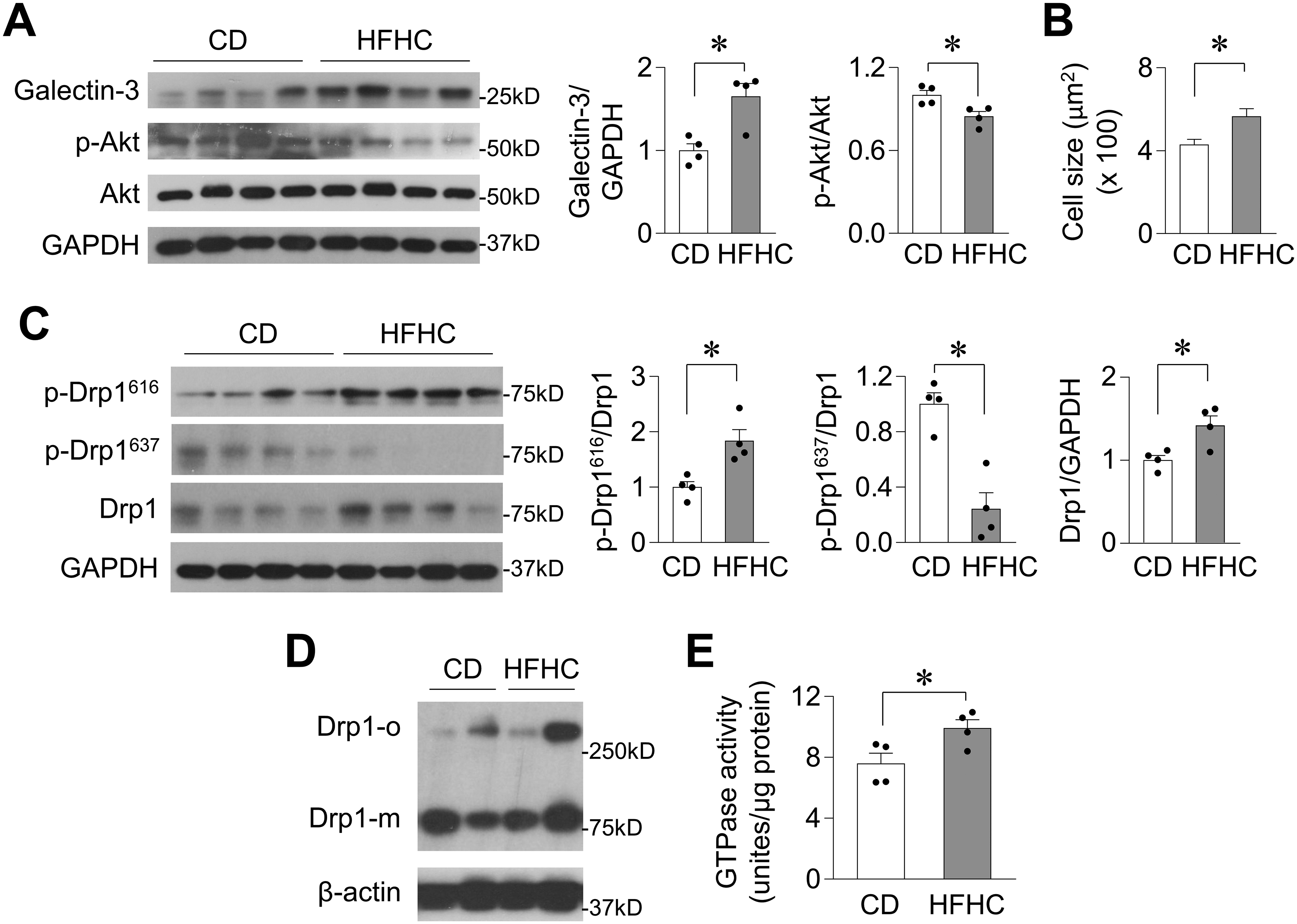
Objective: To investigate the regulation and function of the mitochondrial fission protein Drp1 (dynamin-related protein 1) in lipid overload-induced cardiomyocyte death and heart dysfunction.

Methods and results: Mice fed a high-fat diet (HFD) developed signs of obesity and type II diabetes mellitus, including hyperlipidemia, hyperglycemia, hyperinsulinemia, and hypertension. HFD for 18 weeks also induced heart hypertrophy, fibrosis, myocardial insulin resistance, and cardiomyocyte death. HFD stimulated mitochondrial fission in mouse hearts. Furthermore, HFD increased the protein level, phosphorylation (at the activating serine 616 sites), oligomerization, mitochondrial translocation, and GTPase activity of Drp1 in mouse hearts, indicating that Drp1 was activated. Monkeys fed a diet high in fat and cholesterol for 2.5 years also exhibited myocardial damage and Drp1 activation in the heart. Interestingly, HFD decreased nicotinamide adenine dinucleotide (oxidized) levels and increased Drp1 acetylation in the heart. In adult cardiomyocytes, palmitate increased Drp1 acetylation, phosphorylation, and protein levels, and these increases were abolished by restoration of the decreased nicotinamide adenine dinucleotide (oxidized) level. Proteomics analysis and in vitro screening revealed that Drp1 acetylation at lysine 642 (K642) was increased by HFD in mouse hearts and by palmitate incubation in cardiomyocytes. The nonacetylated Drp1 mutation (K642R) attenuated palmitate-induced Drp1 activation, its interaction with voltage-dependent anion channel 1, mitochondrial fission, contractile dysfunction, and cardiomyocyte death.

Conclusions: These findings uncover a novel mechanism that contributes to lipid overload-induced heart hypertrophy and dysfunction. Excessive lipid supply created an intracellular environment that facilitated Drp1 acetylation, which, in turn, increased its activity and mitochondrial translocation, resulting in cardiomyocyte dysfunction and death. Thus, Drp1 may be a critical mediator of lipid overload-induced heart dysfunction as well as a potential target for therapy.

Keywords: acetylation; diabetes mellitus; dynamins; heart; mitochondria.


Figures

Figure 1.
Figure 1.. High-fat diet induced heart dysfunction in mice.
A, Palmitate (C16:0) and oleate (C18:1) levels in plasma (left) or heart samples (right) from mice fed regular rodent chow (RC) or high-fat diet (HFD) for 18 weeks. N=10 for each group. B, Plasma insulin levels (left, N=5–6) or diastolic and systolic blood pressure (right, N=9–12) in mice fed different diets for 18 weeks. C, Quantification of heart weight (HW) over tibia length (TL) ratio (N=5 for each group), Left ventricle (LV) mass (N=4–6), and plasma BNP levels (N=6) in mice fed different diets for 18 weeks. D, Fractional shortening (FS) in mice fed different diets. N=6. E, Western blotting images and summarized data showing phospho-Akt (Serine 473), total Akt, galectin-3, cleaved caspase 9, and LC3II/I protein levels in the hearts of mice fed different diets for 18 weeks. N=3–6. F, Electron microscopic images and quantification of mitochondrion size in mouse hearts. N=361–420 mitochondria from 5 mice for each group. Scale bar = 1 μm. *: P<0.05.
Figure 2.
Figure 2.. High-fat diet activated Drp1 in mouse hearts.
A, Western blotting images and summarized data showing Drp1 and Opa1 protein levels in whole heart (left) and Drp1 protein levels in mitochondrial or cytosolic fractions (right) from mice fed different diets for 18 weeks. N=5. B-C, Western blotting images and summarized data showing Drp1 phosphorylation at serine 616 (p-Drp1616) or serine 637 (p-Drp1637) in whole heart (B) or subcellular fractions (C) of mouse hearts. N=6. D-E, Western blotting images and summarized data showing Drp1 oligomerization (Drp1-o) in whole heart (D) or subcellular fractions (E) of mouse hearts. N=6. F, The GTPase activity of Drp1 in mouse hearts. N=3. *: P<0.05.
Figure 3.
Figure 3.. High-fat and high-cholesterol diet damaged myocardium and activated Drp1 in monkey hearts.
A, Western blotting images and summarized data showing galectin-3, Akt phosphorylation (p-Akt), Akt, and GAPDH levels in the heart of monkeys fed a control diet (CD) or a high-fat and high-cholesterol (HFHC) diet for 2.5 years. N=4 per group. B, Summarized data showing cross-sectional area of cardiomyocytes in the left ventricular free wall of monkey hearts. N=140–156 cells from 4 monkeys in each group. C-D, Western blotting images and summarized data showing Drp1 phosphorylation and protein level (C, N=4) and Drp1 oligomerization (D, N=2) in monkey heart samples. E, The GTPase activity of Drp1 in monkey heart samples. N=4. *: P<0.05.
Figure 4.
Figure 4.. High-fat diet created an intracellular environment that promoted Drp1 acetylation.
A, Total NAD+ content (left, N=3–4) and cytosolic free NAD+/NADH ratios (right, N=7) in heart tissues from mice fed different diets for 18 weeks. B, Protein levels of NAMPT in mouse hearts. N=3. C, Acetyl-CoA levels in mouse hearts. N=4. D, Total protein acetylation levels in the heart of mice fed different diets for 18 weeks. N=4. E-G, Western blotting images and summarized data showing Drp1 acetylation in mouse hearts (E), monkey hearts (F), or subcellular fractions of mouse hearts (G). N=4–6. *: P<0.05.
Figure 5.
Figure 5.. Lipid overload activated Drp1 in adult cardiomyocytes.
A, Total intracellular NAD+ content (left, N=3–9) and cytosolic free NAD+/NADH ratios monitored by SoNar indicator (right, N=28–29 cells from 3 rats) in adult cardiomyocytes incubated with palmitate (Pal, 0.3 mmol/L) and with pretreatment of NAD+ precursor NMN (500 μmol/L). B, Effects of palmitate (0.3 mmol/L, 24 hr) on the protein levels of NAMPT (left), Sirt1, and acetyl-CoA synthetase (ac-CoA syn) (right) in adult cardiomyocytes. N=3 rats. C, Dose- (left, N=6) and time-dependent (middle, N=4) effects of palmitate on Drp1 acetylation with or without NAD+ precursor (NR, 1 mmol/L) and the effect of overexpressing NAMPT on palmitate-induced Drp1 acetylation (right, N=3) in adult cardiomyocytes. D, Western blotting images and summarized data showing time-dependent effects of palmitate (0.3 mmol/L) on Drp1 and Opa1 protein levels in adult cardiomyocytes. N=3–9 rats. E, Western blotting images showing time- and dose-dependent effects of palmitate on Drp1 S616 phosphorylation in adult cardiomyocytes and with pretreatment of NAD+ precursor (NR, 1 mmol/L). N=5–6 rats. F, Western blotting images and summarized data showing Drp1 oligomerization (Drp1-o) in adult cardiomyocytes. N=6 rats. G, Representative confocal images and summarized data showing GFP-Drp1 puncta in live adult cardiomyocytes. N=109–177 puncta from 3–4 rats. Scale bars = 20 or 5 μm for the whole cell or enlarged images, respectively. *: P<0.05.
Figure 6.
Figure 6.. Lipid overload increased Drp1 acetylation at lysine 642.
A, Western blotting images showing acetylation of purified Drp1 after incubation with acetyl-CoA (upper) and scheme showing the location of 10 lysine residues in Drp1 that can be acetylated in vitro (lower). The domains are GTPase domain (GTP), middle domain (M), B domain (B), and GTPase effector domain (GED). B, Mass spectrometry analysis of Drp1 acetylation showing the tandem mass spectrum of a Drp1 fragment containing the acetylated K642 (increased by 42 Da) in the heart samples of HFD-fed mice. C-E, Western blotting images showing the effects of palmitate (Pal, 0.3 mmol/L, 24 hr) on the protein levels (C), acetylation (D), and oligomerization (E) of overexpressed wild type (WT) or mutations of Drp1 (non-tagged) in HEK293 cells. N=5–6. F, Summarized data showing the effects of palmitate on mitochondrion size in HEK293 cells overexpressing various Drp1 mutations (non-tagged). N=16564–30141 mitochondria from 4 independent experiments. *: P<0.05.
Figure 7.
Figure 7.. Preventing Drp1 acetylation at K642 blocked palmitate-induced Drp1 activation and fission.
A, Western blotting images and summarized data showing the degradation of GFP-tagged Drp1 (WT) or Drp1 K642R mutation (K642R) with or without palmitate and cycloheximide (CHX, 25 μg/ml, 24 hr) incubation. N=5 rats. B-D, Western blotting images and summarized data showing the effects of WT or K642R on palmitate-induced Drp1 acetylation (B), S616 phosphorylation (C), and oligomerization (D). N=5 rats. E, Representative confocal images and summarized data showing GFP puncta and mitochondrial morphology in adult cardiomyocytes overexpressing WT or K642R and in the presence of palmitate. N=763–1221 mitochondria or 20839–39002 GFP puncta in 18–24 cells from 3–4 rats in each group. Scale bars = 20 or 5 μm for the whole cell or enlarged images, respectively. F, Representative confocal images showing mitochondrial morphology in Drp1 knockout (KO) MEF cells overexpressing WT or K642R and in the presence of palmitate. N=16 cells in each group from 3 independent experiments. G, The GTPase activity of Drp1 in adult cardiomyocytes overexpressing WT or K642R. N=3 rats. *: P<0.05.
Figure 8.
Figure 8.. Acetylation at K642 increased Drp1 and VDAC1 interaction and mediated lipid overload-induced cardiomyocyte dysfunction.
A, Western blotting images and summarized data showing Drp1 and VDAC1 interactions in the heart of mice fed different diets for 18 weeks. N=3 rats. B, Western blotting image and summarized data showing the interactions between VDAC1 and WT Drp1 or VDAC1 and Drp1 K642R in adult cardiomyocytes in the presence of palmitate. N=3 rats. C, Representative original or normalized traces (upper) and summarized data (lower) showing the effects of palmitate (0.3 mmol/L, 24 hr) on electric pacing-induced cardiomyocyte contraction (cell shortening) and relaxation (T50%) and with overexpression of K642R or pretreatment with VBit4 (10 μmol/L). N=30–43 cells in each group from 3–4 rats. D-F, Effects of palmitate on mPTP time (D), cell death (E), cleaved caspase 9 (F), and LC3II/I ratios (F) in adult cardiomyocytes and with overexpression of K642R or pretreatment with VBit4. N=3–4 rats. *: P<0.05.

References

    1. Ortega FB, Lavie CJ, Blair SN. Obesity and cardiovascular disease. Circ Res. 2016;118:1752–1770 - PubMed
    1. Beckman JA, Creager MA. Vascular complications of diabetes. Circ Res. 2016;118:1771–1785 - PubMed
    1. Kenny HC, Abel ED. Heart failure in type 2 diabetes mellitus. Circ Res. 2019;124:121–141 - PMC - PubMed
    1. Reaven GM, Lithell H, Landsberg L. Hypertension and associated metabolic abnormalities--the role of insulin resistance and the sympathoadrenal system. N Engl J Med. 1996;334:374–381 - PubMed
    1. Wende AR, Abel ED. Lipotoxicity in the heart. Biochim Biophys Acta. 2010;1801:311–319 - PMC - PubMed

Publication types

MeSH terms