Review
. 2020 Jan 2;40(1):22-36.
doi: 10.1523/JNEUROSCI.0736-19.2019.

Generation, Coordination, and Evolution of Neural Circuits for Vocal Communication

Affiliations
Review

Generation, Coordination, and Evolution of Neural Circuits for Vocal Communication

Darcy B Kelley et al. J Neurosci. .

Abstract

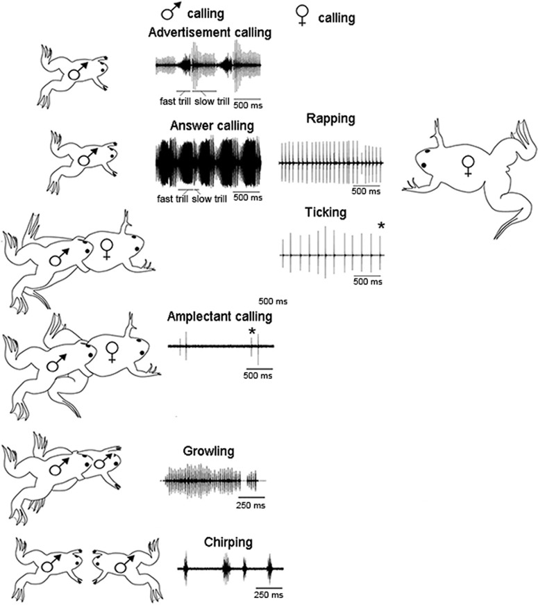
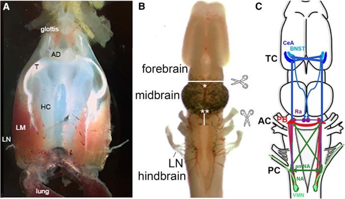
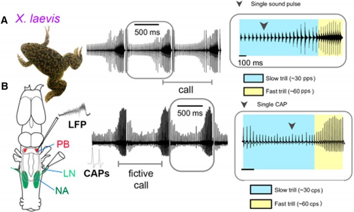
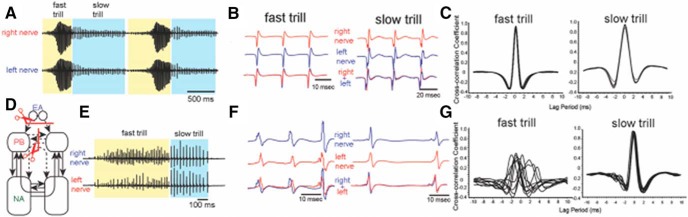
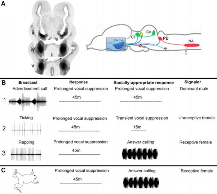
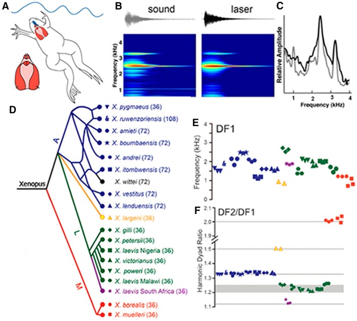
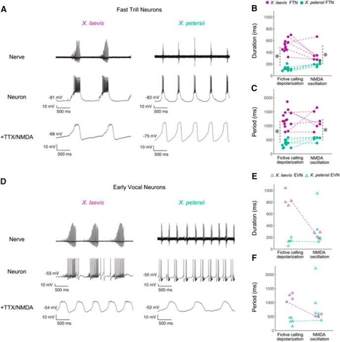
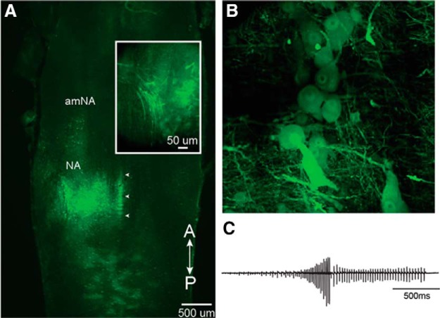
In many species, vocal communication is essential for coordinating social behaviors including courtship, mating, parenting, rivalry, and alarm signaling. Effective communication requires accurate production, detection, and classification of signals, as well as selection of socially appropriate responses. Understanding how signals are generated and how acoustic signals are perceived is key to understanding the neurobiology of social behaviors. Here we review our long-standing research program focused on Xenopus, a frog genus which has provided valuable insights into the mechanisms and evolution of vertebrate social behaviors. In Xenopus laevis, vocal signals differ between the sexes, through development, and across the genus, reflecting evolutionary divergence in sensory and motor circuits that can be interrogated mechanistically. Using two ex vivo preparations, the isolated brain and vocal organ, we have identified essential components of the vocal production system: the sexually differentiated larynx at the periphery, and the hindbrain vocal central pattern generator (CPG) centrally, that produce sex- and species-characteristic sound pulse frequencies and temporal patterns, respectively. Within the hindbrain, we have described how intrinsic membrane properties of neurons in the vocal CPG generate species-specific vocal patterns, how vocal nuclei are connected to generate vocal patterns, as well as the roles of neurotransmitters and neuromodulators in activating the circuit. For sensorimotor integration, we identified a key forebrain node that links auditory and vocal production circuits to match socially appropriate vocal responses to acoustic features of male and female calls. The availability of a well supported phylogeny as well as reference genomes from several species now support analysis of the genetic architecture and the evolutionary divergence of neural circuits for vocal communication. Xenopus thus provides a vertebrate model in which to study vocal communication at many levels, from physiology, to behavior, and from development to evolution. As one of the most comprehensively studied phylogenetic groups within vertebrate vocal communication systems, Xenopus provides insights that can inform social communication across phyla.

Keywords: CPG; duets; hindbrain; neuroendocrine; parabrachial; song.


Figures

Figure 1.
Figure 1.
The vocal repertoire of X. laevis is sexually differentiated and specific to social context. The male advertisement call consists of individual sound pulses* at fast (60 pps) and slow (30 pps) rates. Males produce advertisement calls when alone or with conspecifics. When oviposition is imminent, females produce a rapid (16 pps) series of sound pulses known as rapping. Rapping serves as an acoustic aphrodisiac and stimulates male answer calling. When clasped, sexually unreceptive females extend their hind legs and produce the slower ticking call (6 pps). A male clasping another frog, male or female, produces the amplectant call. A clasped male responds with growling. During the social interactions that precede one male vocally suppressing another, males produce the chirping call. Modified from Zornik and Kelley, 2011. pps: pulses per second.
Figure 2.
Figure 2.
A, The ex vivo larynx, dorsal view; anterior is up. The larynx communicates with the oral cavity via the glottis anteriorly and with the lungs posteriorly. The sound producing elements are the arytenoid disks (ADs), which connect via a tendon (T) to intrinsic laryngeal muscles (LM), which wrap around the hyaline cartilage (HC). When the laryngeal nerves (LNs) are stimulated simultaneously, the muscles contract and, provided a critical velocity is attained, the ADs separate and a sound pulse is produced. B, The ex vivo brain viewed from the dorsal surface, anterior is up. Locations of transections discussed are indicated by scissors icons, white lines, and asterisks. C, A schematic diagram of brain regions that contribute to vocal production including the CeA and BNST in the forebrain (blue), the PB (red) and amNA (green) in the hindbrain, and the raphe nucleus (purple), as well as extensive, ipsilateral and contralateral, usually reciprocal, connections between each nucleus. TC, Telencephalic commissure; AC, anterior commissure; PC, posterior commissure.
Figure 3.
Figure 3.
Actual (A) and fictive (B) advertisement calling in Xenopus laevis. Compound action potentials (CAPs) recorded from the laryngeal nerve correspond to pattern of actual advertisement calls. A LFP that coincides with fast trill CAPs from the laryngeal nerve (LN), can be recorded extracellularly from the PB. Modified from Barkan et al. (2017, .
Figure 4.
Figure 4.
Transecting both the anterior commissure (Fig. 2C) and forebrain input (including the EA:CeA plus BNST) to PB disrupts synchrony of fictive fast but not slow trills. A, Recordings from the left and right laryngeal nerves in an intact male brain. B, Enlarged views of left (blue) and right (red) nerve recordings during fictive fast and slow trills; traces from left and right nerve overlap. C, Example cross-correlation between the left and right nerve during fictive fast and slow trills calculated by a sliding a recording of a single compound action potential from one nerve over the other. Peak cross-correlation coefficients are centered around zero, indicating that the two nerves are active simultaneously. D, Bilateral forebrain and anterior commissure input to the PB were removed. Transection is indicated by a red line with scissors, transected projections by dotted arrows, and intact projections by solid arrows. E, Recordings from the left (top trace) and right (bottom trace) laryngeal nerves in an intact male brain. F, Enlarged views of left (red) and right (blue) nerve recordings during fictive fast and slow trills in a transected brain. The bottom trace shows the overlay of the left and right nerve recordings. G, A cross-correlation between the left and right nerve during fast and slow trills generated by a double-transected brain. The peak cross-correlation coefficient for slow trills is centered around zero, as in the intact brain, but for fast trills correlation coefficients are variable, indicating that the left and right laryngeal nerves are activated simultaneously during slow trills, but asynchronously during fast trills. Modified from Yamaguchi et al. (2017).
Figure 5.
Figure 5.
Damage to the CeA of males results in socially inappropriate responses to female calls. Effects of different calls were assessed in males with lesions of the EA (CeA and BNST). Males called spontaneously after CeA lesions so were not mute. A, Lesions of the EA are shown in transverse sections through the forebrain (Hall et al., 2013) and schematically in a sagittal view. Auditory nuclei in green, components of the vocal CPG in red. B1, Lesioned males responded to broadcasts of male advertisement calls with prolonged vocal suppression, the socially appropriate response, so lesioned males were not deaf. B2, Lesioned males respond to ticking with prolonged rather than socially-appropriate transient vocal suppression. B3, Lesioned males responded to broadcasts of rapping with prolonged vocal suppression rather than answer calling. C, Even when paired with a rapping, receptive female, CeA-lesioned males exhibit prolonged vocal suppression.
Figure 6.
Figure 6.
A, Advertisement calls were recorded underwater from vocalizing males or from the ex vivo larynx, at left. Note the relative sizes of the brain (blue) and larynx (red). B, A single sound pulse. Acoustic (gray) or laser inferometry (black) recordings reveal two dominant frequencies (C). D, Phylogenetic representation of species and populations from which advertisement calls were recorded, color-coded by clade (A, blue; L, green and purple; M, red and black). Ploidy levels for each species are in parentheses: 36 = tetraploid. Symbols correspond to individuals of each species in E and F. Updated according to Evans et al. (2019) for X. fraseri. E, Although both the lower DF1 and higher (data not shown) DF2 overlap across species, their ratios (F) are species-group. DF2–DF1 ratios and their harmonic intervals: 2.0 (octave), 1.5 (perfect 5th), 1.34 (perfect 4th), and 1.22,(minor 3rd), 1.25 (major 3rd). For X. itombuensis audio recording, see Movie 1. Modified from Kwong-Brown et al. (2019).
Figure 7.
Figure 7.
The PB includes two classes of intrinsically, rhythmic neurons: early vocal neurons (EVNs) and fast trill neurons (FTNs). A, D, Laryngeal nerve and intracellular recordings in PB from X. laevis and X. petersii. The duration and period of FTN neuron depolarization and NMDA-induced oscillation (B, C), but not EVN neuron depolarization and membrane oscillation (E, F), match species-specific temporal features of calls. Asterisks indicated significant differences between species. Reproduced from Barkan et al. (2018).
Figure 8.
Figure 8.
Ex vivo brains injected with vesicular stomatitis virus (VSV) into NA generate fictive advertisement calls. A, Unilateral injection into NA of recombinant VSV encoding its own glycoprotein [rVSV(VSV-G)]. White arrowheads indicate the midline. Inset, NA expressing Venus 2 at 48 h post-injection. Note labeled axons in the laryngeal nerve at left. B, Reporter gene expression in the somata of vocal motor neurons. C, Fictive advertisement call in response to the application of serotonin elicited from the brain illustrated in A. Modified from Yamaguchi et al. (2018).

References

    1. Albersheim-Carter J, Blubaum A, Ballagh IH, Missaghi K, Siuda ER, McMurray G, Bass AH, Dubuc R, Kelley DB, Schmidt MF, Wilson RJ, Gray PA (2016) Testing the evolutionary conservation of vocal motoneurons in vertebrates. Respir Physiol Neurobiol 224:2–10. 10.1016/j.resp.2015.06.010 - DOI - PMC - PubMed
    1. Baertsch NA, Baertsch HC, Ramirez JM (2018) The interdependence of excitation and inhibition for the control of dynamic breathing rhythms. Nat Commun 9:843. 10.1038/s41467-018-03223-x - DOI - PMC - PubMed
    1. Barkan CL, Zornik E (2019) Feedback to the future: motor neuron contributions to central pattern generator function. J Exp Biol 222:jeb193318. 10.1242/jeb.193318 - DOI - PMC - PubMed
    1. Barkan CL, Zornik E, Kelley DB (2017) Evolution of vocal patterns: tuning hindbrain circuits during species divergence. J Exp Biol 220:856–867. 10.1242/jeb.146845 - DOI - PMC - PubMed
    1. Barkan CL, Kelley DB, Zornik E (2018) Premotor neuron divergence reflects vocal evolution. J Neurosci 38:5325–5337. 10.1523/JNEUROSCI.0089-18.2018 - DOI - PMC - PubMed

Publication types

MeSH terms

Substances